Review
. 2024 May 26;25(11):5779.
doi: 10.3390/ijms25115779.

Structural Features Influencing the Bioactive Conformation of Angiotensin II and Angiotensin A: Relationship between Receptor Desensitization, Addiction, and the Blood-Brain Barrier

Affiliations
Review

Structural Features Influencing the Bioactive Conformation of Angiotensin II and Angiotensin A: Relationship between Receptor Desensitization, Addiction, and the Blood-Brain Barrier

Graham J Moore et al. Int J Mol Sci. .

Abstract

The N-terminal portion of the octapeptide angiotensin II (DRVYIHPF; AngII), a vasopressor peptide that favorably binds to, and activates, AngII type 1 receptor (AT1R), has an important role in maintaining bioactive conformation. It involves all three charged groups, namely (i) the N-terminal amino group cation, (ii) the Asp sidechain anion and (iii) the Arg guanidino cation. Neutralization of any one of these three charged groups results in a substantial reduction (<5%) in bioactivity, implicating a specialized function for this cluster. In contrast, angiotensin A (ARVYIHPF; AngA) has reduced bioactivity at AT1R; however, replacement of Asp in AngII with sarcosine (N-methyl-glycine) not only restores bioactivity but increases the activity of agonist, antagonist, and inverse agonist analogues. A bend produced at the N-terminus by the introduction of the secondary amino acid sarcosine is thought to realign the functional groups that chaperone the C-terminal portion of AngII, allowing transfer of the negative charge originating at the C-terminus to be transferred to the Tyr hydroxyl-forming tyrosinate anion, which is required to activate the receptor and desensitizes the receptor (tachyphylaxis). Peptide (sarilesin) and nonpeptide (sartans) moieties, which are long-acting inverse agonists, appear to desensitize the receptor by a mechanism analogous to tachyphylaxis. Sartans/bisartans were found to bind to alpha adrenergic receptors resulting in structure-dependent desensitization or resensitization. These considerations have provided information on the mechanisms of receptor desensitization/tolerance and insights into possible avenues for treating addiction. In this regard sartans, which appear to cross the blood-brain barrier more readily than bisartans, are the preferred drug candidates.

Keywords: G-protein coupled receptor; addiction; angiotensin II; arginine; bisartan; blood–brain barrier; conformation of angiotensin; coronavirus disease 2019; receptor desensitization.


Conflict of interest statement

A.Z. co-owns Zultek Engineering, the provider of product OB16 organ bath systems. Authors J.M.M. and K.K. were employed by NewDrug and author G.J.M. by Pepmetics Inc. The remaining authors declare that the research was conducted in the absence of any commercial or financial relationships that could be construed as a potential conflict of interest.

Figures

Figure 1
Figure 1
Schematic diagram of the brain RAS, depicting the various peptides, enzymes and receptors involved and the subsequent effect on specific neurological functions when activated by their ligands [44,45,46]. Angiotensinogen is first converted to AngI by renin and its precursor, prorenin. AngI can be metabolized to AngII by ACE and chymase, converted to Ang(1–9) by ACE2 or catalyzed into Ang(1–7) by neprilysin [31,45]. AngII can associate with either AT1R or AT2R, resulting in detrimental or homeostatic promoting effects, respectively [31,45]. Furthermore, AngII can then be converted to AngIII by aminopeptidase A (APA), AngA by aspartate decarboxylase (AD) or Ang(1–7) by ACE2 [31,45]. The resulting Ang III peptide is further acted upon by APA and alanyl aminopeptidase to become AngIV, which interacts with AT4R to promote neuroprotective effects [31,45]. Ang(1–9) can be catalyzed by ACE to either directly bind to the Mas-related G-coupled protein receptor (MrgDR) or can be acted upon by AD into alamandine, interacting with the Mas1 oncogene receptor (MasR) [31,45]. Finally, AngA can also be converted to alamandine by ACE2 [26]. Activation of prorenin receptor or AT1R by renin/prorenin and AngII, respectively, results in deleterious events, such as cell death, inflammation, oxidative stress, and vasoconstriction [44,45,46]. In contrast, activation of AT2R, AT4R, MasR and MrgDR induces opposing advantageous effects [44,45,46]. Abbreviations: ACE, angiotensin-converting enzyme; AD, aspartate decarboxylase; Ala, alanine; AngI, angiotensin; AngII, angiotensin II; AngIII, angiotensin III; AngIV, angiotensin IV; APA, aminopeptidase A; APN, alanyl aminopeptidase; Arg, arginine; Asp, aspartic acid; AT1R, angiotensin II type 1 receptor; AT2R, angiotensin II type 2 receptor; AT4R, angiotensin II type 4 receptor; His, histidine; Ile, isoleucine; Leu, leucine; MasR, Mas1 oncogene receptor; MrgDR, Mas-related G-coupled protein receptor; NEP, neprilysin; Phe, phenylalanine; Pro, proline; Ser, serine; PRR, prorenin receptor; RAS, renin–angiotensin system; Thr, threonine; Tyr, tyrosine; Val, valine. Figure made using Biorender.com (access date: 10 March 2024).
Figure 2
Figure 2
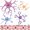
Expression profile of brain RAS components and α1 adrenergic receptors within the BBB, including astrocytes, microglia, neuron, and oligodendrocytes [33,47,48]. While it was once thought that AngII was unable to pass through the BBB due to its size and hydrophobic nature, there is evidence to the contrary. Firstly, in spontaneous hypertensive rats, breakdown of the BBB may increase permeability, allowing for extravasate access of AngII into the hypothalamus [49]; however further research is required to substantiate this claim in humans. Moreover, AT1R-mediated transcytosis of AngII by brain endothelial cells (BECs) of the BBB may allow entry of AngII to regulate autonomic function [50]. Abbreviations: ACE, angiotensin-converting enzyme; AGT, angiotensinogen; Ang, angiotensin; APA, aminopeptidase A; AT1R, angiotensin II type 1 receptor; AT2R, angiotensin II type 2 receptor; AT4R, angiotensin II type 4 receptor; α1AR, adrenergic receptors; BECs, brain endothelial cells; MasR, Mas1 oncogene receptor; MrgDR, Mas-related G-coupled protein receptor; PRR, prorenin receptor; RAS, renin–angiotensin system. Key: red dashed arrow = peptide binds to and activates AT1R; green dashed line = peptide binds to and activates AT2R; orange dashed arrow = peptide binds to MrgDR; purple dashed arrow = peptide binds to and activates MasR; brown dashed arrows = peptide binds to and activates PRR. Figure made using Biorender.com (access date: 10 March 2024).
Figure 3
Figure 3
Two- and three-dimensional models depict intramolecular interactions of angiotensins. (A) The tyrosinate anion, formed by the charge relay system (CRS) (Tyr..His..CO2- of Phe) is stabilized by interaction with the arginine (Arg) guanidino cation, which is maintained in position by the aspartate (Asp) carboxylate anion, depicted bonded between two cations (resulting in bioactivity as charge is transferred to TyrOH). (B) Angiotensin A (AngA) alters the conformation of the N-terminal region, affecting the ability of the Arg cation to effectively chaperone the CRS (resulting in weak activity as charge transfer is only partial). (C) [Sar1] AngII creates a bend at the N-terminus restoring the position of the Arg guanidino cation, enabling it, together with the sarcosine (Sar) amino cation, to chaperone the CRS (enhancing bioactivity and functioning as a superagonist). Abbreviations: AngII, angiotensin II; C, carbon; H, hydrogen; His, histidine; Ile, isoleucine; N, nitrogen; O, oxygen; Phe, phenylalanine; Pro, proline; Tyr, tyrosine; Val, valine. Key: blue = nitrogen atoms; red = oxygen atom; grey= carbon atoms; white = hydrogen atoms.
Figure 4
Figure 4
Receptor interactions of the cardiovascular effects of agonist and inverse agonist (desensitizing) effects at AT1R. The figure illustrates the dynamic interaction between AngII and AT1R under different conditions [82,83,84,85]. At baseline, AngII binds to AT1R in its resting state. At submaximal doses (nH > 1 < 2), AngII forms an activated state with the alpha, beta, and gamma G-protein subunits, leading to endothelial dysfunction, the pathogenesis of cardiovascular diseases (CVD), vasoconstriction, cardiac remodeling, pro-oxidative stress, and inflammation. Conversely, at supramaximal doses (nH < 1), inverse agonists induce an inverted state of the receptor. This results in an increase in nitric oxide (NO) and cardiac performance, decreased cellular stress, cardioprotection, vasodilation, anti-fibrotic, anti-oxidative, and anti-inflammatory effects conditions [82,83,84,85]. Desensitizing or inverse agonists form more stable salt bridges with residue R167 of AT1R, coupling the receptor to different signaling pathways and inducing long-term blockade, as seen with certain ARBs, like candesartan, telmisartan, and the peptide analogue sarilesin. Additionally, Figure 4 suggests a potential mechanism for tachyphylaxis, wherein high concentrations of agonists lead to receptor desensitization by forming stable salt bridges, resulting in a slow reversal process. This mechanism likely serves as a cellular defense mechanism against excessive cell stimulation, particularly in cardiovascular tissues, thereby protecting against the noxious effects of AngII. Abbreviations: P, phosphorylation. Key: ↑ = increases; ↓ = decreases. Figure created with Biorender.com (access date: 10 March 2024).
Figure 5
Figure 5
Structures of the ligands which were used for docking studies to the X-ray crystal structure of the alpha receptor are shown. Abbreviations: BisA, 4-Butyl-N,N0-bis([[20-(2H-tetrazol-5-yl)]biphenyl-4-yl]) methylimidazolium bromide; BisB, 4-Butyl-2-hydroxymethyl-N,N0-bis([20-(2H-tetrazol-5-yl)- biphenyl-4-yl])methyimidazolium bromide; BisC, 2-Butyl-4-chloro-5-hydroxymethyl-N,N0-bis([20-(2H-tetrazol- 5-yl)biphenyl-4-yl]methyl)imidazolium bromide; BisD, 2-Butyl-N,N0-bis([20-(2H-tetrazol-5-yl)biphenyl-4-yl]methyl) imidazolium bromide; Cl, chlorine; DIZE, diminazene aceturate; exp3174, losartan carboxylic acid; H, hydrogen; N, nitrogen; O, oxygen; S, sulfur. Key: green = chlorine; grey = hydrogen; blue = nitrogen; red = oxygen; yellow = sulfur.
Figure 6
Figure 6
(A) Docking results obtained for 29 selected ligands, including various sartans (blue bars) and known inverse agonists to an alpha-2C-adrenergic receptor (green and orange bars), belonging to the GPCR class. Ligand docking was performed against the energy-minimized (AMBER14) alpha-2c-adrenergic receptor, PDB 6KUW, using AutoDock VINA with 900 runs per ligand with assigned AMBER14 atomic point charges and dihedral barriers [28]. Of the 29 ligands evaluated, the anionic bisartan ACC519TT exhibited the strongest docking energy (12.5 kcal/mol). ACC519TT outperformed the opioid-like ligand “6kuwLig” (compound RS79948:(8~(a)~(R),12~(a)~(S),13~(a)~(R))-12-orange bars) (accessed on 11 March, 2024) designed as a specific inhibitor of the 6KUW receptor. (B) Structure of the 6KUW opioid receptor with docked bisartan ACC519TT (yellow carbon atoms) embedded in the cell surface domain. The yellow shaded area approximates the docking region of interest. Residues shown in ethylsulfonyl-3-methoxy-5,6,8,8~(a),9,10,11,12~(a),13,13~(a)-decahydroisoquinolino [2,1-g][1,6]naphthyridine; https://www.rcsb.org/structure/6KUW; “stick” rendering indicate the locations of arginine residues for 6KUW. (C) Illustrates the 3D rendition of the docked ligand ACC519TT, while (D) presents the 2D interaction diagram calculated using PoseView-2D, ZBH-Center for Bioinformatics: https://proteins.plus/) (accessed on 11 March 2024). Principle binding interactions included non-covalent pi–pi resonances (red lines in (C) and cyan dashed lines in (D)) of both anionic tetrazole groups with proximal tyrosine residues (Tyr402 and Tyr405) and phenylalanine (Phe398). Hydrophobic interactions (green lines) were also heavily represented in ACC519TT binding. (E) Superposition of the PDB 6KUW X-ray crystallographic structure of the co-crystallized “6kuwLig” ligand (blue carbon atoms, as spheres) against docked ligand “6kuwLig” (maroon colored carbon atoms, as spheres). The superimposed molecules exhibited an RMSD of 1.0339 Angstroms, indicating that VINA algorithms were able to reproduce a high-quality experimental pose for this ligand. Abbreviations: ACC519TT, benzimidazole-N-biphenyl tetrazole; ACC519[1], benzimidazole-N-biphenyl tetrazole; Asn, asparagine; Azil, azilsartan; BisA, 4-Butyl-N,N0-bis([[20-(2H-tetrazol-5-yl)]biphenyl-4-yl]) methylimidazolium bromide; BisB, 4-Butyl-2-hydroxymethyl-N,N0-bis([20-(2H-tetrazol-5-yl)- biphenyl-4-yl])methyimidazolium bromide; BisC, 2-Butyl-4-chloro-5-hydroxymethyl-N,N0-bis([20-(2H-tetrazol- 5-yl)biphenyl-4-yl]methyl)imidazolium bromide; BisD, 2-Butyl-N,N0-bis([20-(2H-tetrazol-5-yl)biphenyl-4-yl]methyl) imidazolium bromide; Cande, candesartan; Dize, diminazene aceturate; Epro, eprosartan; exp3174, losartan carboxylic acid; Glu, glutamic acid; Gly, glycine; GPCRs, G-protein coupled receptors; Ireb, irbesartan; Kd, dissociation constants; Leu, leucine; Lo, losartan; norepineph, norepinephrine; PDB, Protein Data Bank; Phe, phenylalanine; phenyleph, phenylephrine; RMSD, root mean standard deviation; Ser, serine; Telmi, telmisartan; Tyr, tyrosine; Val, valsartan (7A) or valine (7C); Å, angstrom.
Figure 7
Figure 7
Effects of ACC519T and candesartan on phenylephrine-induced contraction of rabbit iliac arteries. Pretreatment with ACC519T significantly augmented contraction responses to phenylephrine (mean ± standard error of the mean (SEM)) (* p < 0.05, **** p < 0.0001); however, no differences in contraction abilities were observed in rings treated with candesartan (mean ± SEM). Abbreviations: ACC519, benzimidazole-N-biphenyl tetrazole; PE, phenylephrine.
Figure 8
Figure 8
(A) 6KUW A chain showing predicted allosteric pockets (green shaded areas). Allosteric sites were calculated using Passer: Protein Allosteric Sites Server (https://passer.smu.edu/; access date: 5 March 2024). Docking was performed against allosteric site 1 for both 6KUW and 7B6W. (B) Binding energies of different drugs competed for the same site on the alpha receptors. Abbreviations: Allo, allosteric; Cande, candesartan; H, hydrogen; Kd, dissociation constants; Metamphet, methamphetamine; N, nitrogen; Norepi, norepinephrine; O, oxygen; Phenyleph, phenylephrine.

Similar articles

Cited by

References

    1. Fluyau D., Hashmi M.F., Charlton T.E. Drug Addiction. StatPearls; Treasure Island, FL, USA: 2024. - PubMed
    1. Lopez-Leon S., Gonzalez-Giraldo Y., Wegman-Ostrosky T., Forero D.A. Molecular genetics of substance use disorders: An umbrella review. Neurosci. Biobehav. Rev. 2021;124:358–369. doi: 10.1016/j.neubiorev.2021.01.019. - DOI - PubMed
    1. Ceceli A.O., Bradberry C.W., Goldstein R.Z. The neurobiology of drug addiction: Cross-species insights into the dysfunction and recovery of the prefrontal cortex. Neuropsychopharmacology. 2022;47:276–291. doi: 10.1038/s41386-021-01153-9. - DOI - PMC - PubMed
    1. Cheron J., Kerchove d’Exaerde A.d. Drug addiction: From bench to bedside. Transl. Psychiatry. 2021;11:424. doi: 10.1038/s41398-021-01542-0. - DOI - PMC - PubMed
    1. Hossain M.K., Davidson M., Kypreos E., Feehan J., Muir J.A., Nurgali K., Apostolopoulos V. Immunotherapies for the treatment of drug addiction. Vaccines. 2022;10:1778. doi: 10.3390/vaccines10111778. - DOI - PMC - PubMed

MeSH terms

Substances

LinkOut - more resources