. 2024 May 28;25(11):5879.
doi: 10.3390/ijms25115879.

A Pilot Study on Circulating, Cellular, and Tissue Biomarkers in Osteosarcopenic Patients

Affiliations

A Pilot Study on Circulating, Cellular, and Tissue Biomarkers in Osteosarcopenic Patients

Francesca Salamanna et al. Int J Mol Sci. .

Abstract

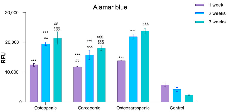
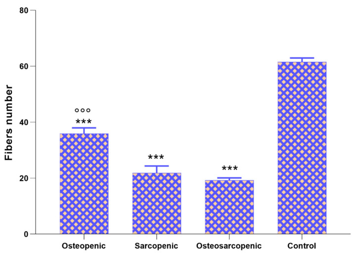
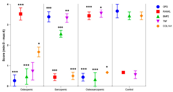
Aging comes with the loss of muscle and bone mass, leading to a condition known as osteosarcopenia. Circulating, cellular, and tissue biomarkers research for osteosarcopenia is relatively scarce and, currently, no established biomarkers exist. Here we find that osteosarcopenic patients exhibited elevated basophils and TNFα levels, along with decreased aPPT, PT/INR, IL15, alpha-Klotho, DHEA-S, and FGF-2 expression and distinctive bone and muscle tissue micro-architecture and biomarker expressions. They also displayed an increase in osteoclast precursors with a concomitant imbalance towards spontaneous osteoclastogenesis. Similarities were noted with osteopenic and sarcopenic patients, including a lower neutrophil percentage and altered cytokine expression. A linear discriminant analysis (LDA) on models based on selected biomarkers showed a classification accuracy in the range of 61-78%. Collectively, our data provide compelling evidence for novel biomarkers for osteosarcopenia that may hold potential as diagnostic tools to promote healthy aging.

Keywords: aging; biomarkers; osteopenia; osteosarcopenia; sarcopenia.


Conflict of interest statement

The authors declare no conflicts of interest.

Figures

Figure 1
Figure 1
Percentage of drug class administered to patients in polypharmacy (a) and comorbidities identified for patients included in the study (b).
Figure 2
Figure 2
Bar plots of the serum levels of selected markers: (a) TNF-α, (b) IL-6, (c) IL-15, (d) IL-1β, (e) IL-33, (f) IL-10, (g) Alpha-KLOTHO, (h) DEHA-S, (i) ST2, (j) FGF2, (k) CNTF, and (l) PGE2 serum levels (Mean ± SD). One-way ANOVA followed by Dunnett’s test of OP, OS, and SP vs. Control (*) and OP and SP vs. OS (°): 1 symbol, p < 0.05, 2 symbols, p < 0.005, 3 symbols, p < 0.0005.
Figure 3
Figure 3
Bar plots of the PBMCs viability of OP, SP, OS, and Control patients after 1, 2, and 3 weeks of culture (Mean ± SD, n = 3 triplicates). Two-way ANOVA (F = 198.8, p < 0.0005) followed by Dunnett’s test of OP, OS, and SP vs. Control (*, at 1 week; °, at 2 weeks; and §, at 3 weeks) and OP and SP vs. OS (#, at 1 week; ^, at 2 weeks; and $, at 3 weeks): 2 symbols, p < 0.005, 3 symbols, p < 0.0005.
Figure 4
Figure 4
Multinucleated TRAP-positive osteoclasts after 3 weeks of culture. (a) OP, (b) SP, (c) OS, and (d) Control patients. TRAP-positive (dark purple) cells with at least three nuclei were considered osteoclasts. Magnification 20×. (e) Number of multinucleated TRAP-positive osteoclasts (three or more nuclei) evaluated in 10 regions of interest (ROI) after 3 weeks of cell culture at 20× magnification (Mean ± SD). One-way ANOVA followed by Dunnett’s test of OP, OS, and SP vs. Control (*) and OP and SP vs. OS (°): 2 symbols, p < 0.005, 3 symbols, p < 0.0005. Scale bar 100 µm.
Figure 5
Figure 5
Histological findings in bone biopsy from (a) OP, (b) SP, (c) OS, and (d) Control patients. Hematoxylin/Eosin staining. Magnification 8×. Osteoporotic cavities (C), bone marrow spaces (Bm), bone trabeculae (Bt), osteocytes (↑), cement lines (arrowhead), refractile areas (*), eroded areas (E), osteoblasts (Ob), and osteoclast (Oc). Scale bar 300 µm.
Figure 6
Figure 6
Histological findings in muscle biopsies from (a) OP, (b) SP, (c) OS, and (d) Control patients. Nuclei (star), myofibers (MF), perimysium (P). Hematoxylin/Eosin staining. Magnification 20×. Scale bar 200 µm.
Figure 7
Figure 7
Number of muscle fibers in muscle biopsies of OP, SP, OS, and Control patients (Mean ± SD). One-way ANOVA (F = 278, p < 0.0005) followed by Dunnett’s test of OP, SP, and OS, vs. Control (*) and OP and SP vs. OS (°): 3 symbols, p < 0.0005.
Figure 8
Figure 8
Dot plot of the results of De la Torre’s score for OPG, RANKL, BMP2, TNF, and COL1A1 immunostaining of OP, SP, OS, and Control patients (Median ± SE). One-way ANOVA followed by Dunnett’s test of OP, SP, and OS, vs. Control (*) and OP and SP vs. OS (°): 1 symbol, p < 0.05, 2 symbols, p < 0.005, 3 symbols, p < 0.0005.

References

    1. Kanis J.A., Oden A., Johansson H., McCloskey E. Pitfalls in the external validation of FRAX: Response to Bolland et al. Osteoporos. Int. 2013;24:391–392. doi: 10.1007/s00198-012-1985-y. - DOI - PubMed
    1. Chen C.Y., Tseng W.C., Yang Y.H., Chen C.L., Lin L.L., Chen F.P., Wong A.M.K. Calf Circumference as an Optimal Choice of Four Screening Tools for Sarcopenia Among Ethnic Chinese Older Adults in Assisted Living. Clin. Interv. Aging. 2020;15:2415–2422. doi: 10.2147/CIA.S287207. - DOI - PMC - PubMed
    1. Cruz-Jentoft A.J., Bahat G., Bauer J., Boirie Y., Bruyère O., Cederholm T., Cooper C., Landi F., Rolland Y., Sayer A.A., et al. Sarcopenia: Revised European consensus on definition and diagnosis. Age Ageing. 2019;48:601. doi: 10.1093/ageing/afz046. - DOI - PMC - PubMed
    1. Kirk B., Zanker J., Duque G. Osteosarcopenia: Epidemiology, diagnosis, and treatment-facts and numbers. J. Cachexia Sarcopenia Muscle. 2020;11:609–618. doi: 10.1002/jcsm.12567. - DOI - PMC - PubMed
    1. Wang Y.J., Wang Y., Zhan J.K., Tang Z.Y., He J.Y., Tan P., Deng H.Q., Huang W., Liu Y.S. Sarco-Osteoporosis: Prevalence and Association with Frailty in Chinese Community-Dwelling Older Adults. Int. J. Endocrinol. 2015;2015:482940. doi: 10.1155/2015/482940. - DOI - PMC - PubMed

LinkOut - more resources