. 2024 May 28;25(11):5889.
doi: 10.3390/ijms25115889.

Anti-HMGB1 mAb Therapy Reduces Epidural Hematoma Injury

Affiliations

Anti-HMGB1 mAb Therapy Reduces Epidural Hematoma Injury

Shangze Gao et al. Int J Mol Sci. .

Abstract

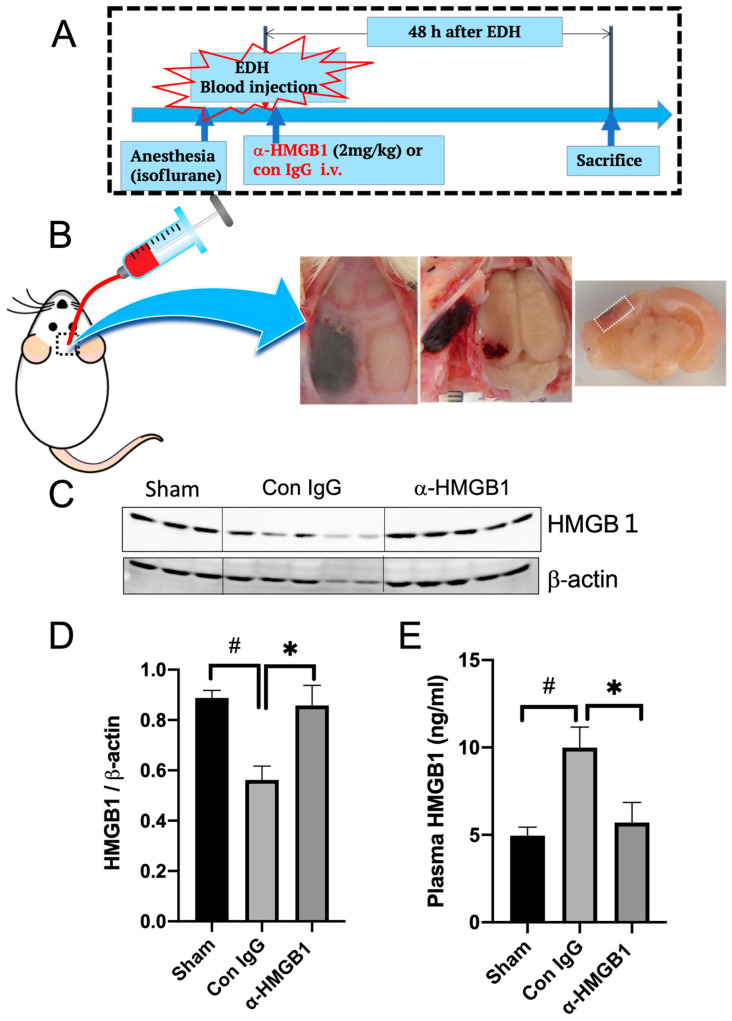
Epidural and subdural hematomas are commonly associated with traumatic brain injury. While surgical removal is the primary intervention for these hematomas, it is also critical to prevent and reduce complications such as post-traumatic epilepsy, which may result from inflammatory responses in the injured brain areas. In the present study, we observed that high mobility group box-1 (HMGB1) decreased in the injured brain area beneath the epidural hematoma (EDH) in rats, concurrent with elevated plasma levels of HMGB1. Anti-HMGB1 monoclonal antibody therapy strongly inhibited both HMGB1 release and the subsequent increase in plasma levels. Moreover, this treatment suppressed the up-regulation of inflammatory cytokines and related molecules such as interleukin-1-beta (IL-1β), tumor necrosis factor-alpha (TNF-α), and inducible nitric oxide synthase (iNOS) in the injured areas. Our in vitro experiments using SH-SY5Y demonstrated that hematoma components-thrombin, heme, and ferrous ion- prompted HMGB1 translocation from the nuclei to the cytoplasm, a process inhibited by the addition of the anti-HMGB1 mAb. These findings suggest that anti-HMGB1 mAb treatment not only inhibits HMGB1 translocation but also curtails inflammation in injured areas, thereby protecting the neural tissue. Thus, anti-HMGB1 mAb therapy could serve as a complementary therapy for an EDH before/after surgery.

Keywords: HMGB1; epidural hematoma; inflammatory response.


Conflict of interest statement

The authors declare no conflict of interest.

Figures

Figure 1
Figure 1
Experimental protocol and changes in HMGB1 levels in the brain and plasma 48 h after the EDH. (A) Experimental protocol of the EDH. (B) Typical appearance picture of the EDH. The brain samples were collected 48 h after the induction of the EDH from the dotted area in (B) and were used for western blotting for HMGB1. (C) The representative results of western blotting are shown. A decrease in HMGB1 levels in the ipsilateral cortex (peri-hematoma) in the control IgG group is shown. (D) Quantitative analyses of western blotting results were performed using NIH Image J software V.1.53, and the results are expressed as mean ± SE of 3–5 rats per group. Differences were considered significant at # p < 0.05 compared to the sham group. * p < 0.05 compared to the control IgG-treated group. (E) Plasma levels of HMGB1 in rats with an EDH were determined by ELISA. Blood samples were collected 48 h after the induction of hemorrhage. The results are expressed as mean ± SE of 3–5 rats per group. # p < 0.05 compared to the sham group. * p < 0.05 compared to the control IgG-treated group.
Figure 2
Figure 2
Effects of the anti-HMGB1 mAb on the expression of inflammation-related molecules and apoptosis 48 h after the EDH. (A) The mRNA expression of TNF-α, iNOS, and IL-1β was measured by quantitative real-time polymerase chain reaction in the cerebral cortex (peri-hematoma) 48 h after the EDH. The primer information refers to previous research [12]. The results are expressed as mean ± SE of 5 rats (A). # p < 0.05 compared to the sham group. * p < 0.05 and compared to the control IgG-treated group. (B) TUNEL staining was performed to reveal apoptotic cells in the ipsilateral cerebral cortex in each group. Scale bar: 50 μm.
Figure 3
Figure 3
Changes in the brain levels of Prxs and heat shock proteins (HSPs) in the brain after the EDH. (A) Prx5 and Prx6 levels in the ipsilateral cerebral cortex (beneath hematoma) were determined 48 h after the EDH by western blotting. (B) The representative results of western blotting are shown. β-actin was used as a reference. Quantitative analyses on western blotting results were performed using NIH Image J software. The results are expressed as mean ± SE of 3–5 rats. ## p < 0.01 compared to the sham group. * p < 0.05 compared to the control IgG-treated group. ns means no significant difference. (C) HSP levels (HSP27, HSP40, HSP60, and HSP90) were determined by western blotting in the ipsilateral cortex (beneath hematoma) 48 h after the EDH. The representative results of western blotting are shown. (D) Quantitative analyses on western blotting results were performed using NIH Image J software. The results are expressed as mean ± SE of 3–5 rats. ### p < 0.001 and # p < 0.05 compared to the sham group. ** p < 0.01 and * p < 0.05 compared to the control IgG-treated group. ns means no significant difference.
Figure 4
Figure 4
Expression of AQP4, PAR1, PAI-1, and MSR1 after the EDH and effects of anti-HMGB1 treatment. (A) The protein levels of AQP4 and PAR-1 in the ipsilateral cerebral cortex (beneath the hematoma) were determined by western blotting 48 h after the EDH. The representative results of western blotting are shown. (B) The quantitative results are expressed as mean ± SE of 3–5 rats. The protein levels of AQP4 and PAR-1 in the ipsilateral cerebral cortex (beneath the hematoma) were determined by western blotting 48 h after the EDH. ## p < 0.01, and ### p < 0.001 compared to the sham group. * p < 0.05 compared to the control IgG-treated group. ns means no significant difference. (C) The protein levels of PAI-1 and MSR-1 in the ipsilateral cerebral cortex (beneath the hematoma) were determined by western blotting 48 h after the EDH. The representative results of western blotting are shown. (D) The quantitative results are expressed as mean ± SE of 3–5 rats. The protein levels of PAI-1 and MSR1 in the ipsilateral cerebral cortex (beneath the hematoma) were determined by western blotting 48 h after the EDH. # p < 0.05, and ### p < 0.001 compared to the sham group. ** p < 0.01, and *** p < 0.01 compared to the control IgG-treated group.
Figure 5
Figure 5
Effects of the anti-HMGB1 mAb on stimulant-induced HMGB1 translocation in neuroblastoma cells in culture. (A) SH-SY5Y neuroblastoma cells were stimulated with hemin (1 mM) for 24 h, and the translocation of HMGB1 was observed immunocytochemically. The anti-HMGB1 mAb (10 μg/mL) or anti-KLH mAb (control) was added to the media immediately before the start of incubation. DAPI was used for nuclear staining. Scale bar: 20 μm. (B) IL-6 and IL-8 levels in the media stimulated with hemin (0.6 mM) were determined 24 h after stimulation in the presence (10 μg/mL) or absence of the anti-HMGB1 mAb. The results are expressed as mean ± SE of triplicate experiments. * p < 0.05 and ** p < 0.01 compared to the non-stimulated group or to the control IgG-treated group, as shown by the lines in the figure. (C) SH-SY5Y neuroblastoma cells were stimulated with FeCl2 (0.6 mM) for 24 h in the presence or absence of the anti-HMGB1 mAb (10 μg/mL), and the translocation of HMGB1 was observed immunocytochemically. Scale bar: 20 μm. (D) SH-SY5Y neuroblastoma cells were stimulated with thrombin (5 U/mL) for 24 h in the presence or absence of the anti-HMGB1 mAb (10 μg/mL), and the translocation of HMGB1 was observed immunocytochemically. Scale bar: 20 μm.
Figure 6
Figure 6
Effects of the anti-HMGB1 mAb on TNF-α-induced HMGB1 translocation in vascular endothelial cells in culture. EA.hy 926 vascular endothelial cells were stimulated with TNF (100 ng/mL) in the presence or absence of the anti-HMGB1 mAb (10 μg/mL) for 6 h, and the translocation of HMGB1 was observed immunocytochemically. DAPI was used for nuclear staining.

References

    1. Aromatario M., Torsello A., D’errico S., Bertozzi G., Sessa F., Cipolloni L., Baldari B. Traumatic Epidural and Subdural Hematoma: Epidemiology, Outcome, and Dating. Medicina. 2021;57:125. doi: 10.3390/medicina57020125. - DOI - PMC - PubMed
    1. Dionyssiotis Y. Analyzing the problem of falls among older people. Int. J. Gen. Med. 2012;5:805–813. doi: 10.2147/ijgm.s32651. - DOI - PMC - PubMed
    1. Tallon J.M., Ackroyd-Stolarz S., Karim S.A., Clarke D.B. The epidemiology of surgically treated acute subdural and epidural hematomas in patients with head injuries: A population-based study. Can. J. Surg. 2008;51:339–345. - PMC - PubMed
    1. Fu L., Liu K., Wake H., Teshigawara K., Yoshino T., Takahashi H., Mori S., Nishibori M. Therapeutic effects of anti-HMGB1 monoclonal antibody on pilocarpine-induced status epilepticus in mice. Sci. Rep. 2017;7:1179. doi: 10.1038/s41598-017-01325-y. - DOI - PMC - PubMed
    1. Nishibori M., Wang D., Ousaka D., Wake H. High Mobility Group Box-1 and Blood–Brain Barrier Disruption. Cells. 2020;9:2650. doi: 10.3390/cells9122650. - DOI - PMC - PubMed

LinkOut - more resources