

Towards Clinical Development of Scandium Radioisotope Complexes for Use in Nuclear Medicine: Encouraging Prospects with the Chelator 1,4,7,10-Tetraazacyclododecane-1,4,7,10-tetraacetic Acid (DOTA) and Its Analogues
- PMID: 38892142
- PMCID: PMC11173192
- DOI: 10.3390/ijms25115954
Towards Clinical Development of Scandium Radioisotope Complexes for Use in Nuclear Medicine: Encouraging Prospects with the Chelator 1,4,7,10-Tetraazacyclododecane-1,4,7,10-tetraacetic Acid (DOTA) and Its Analogues
Abstract
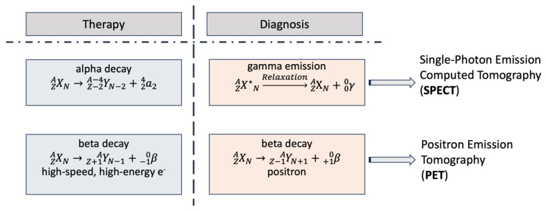
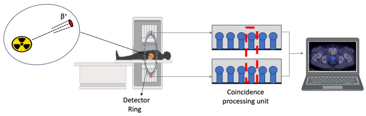
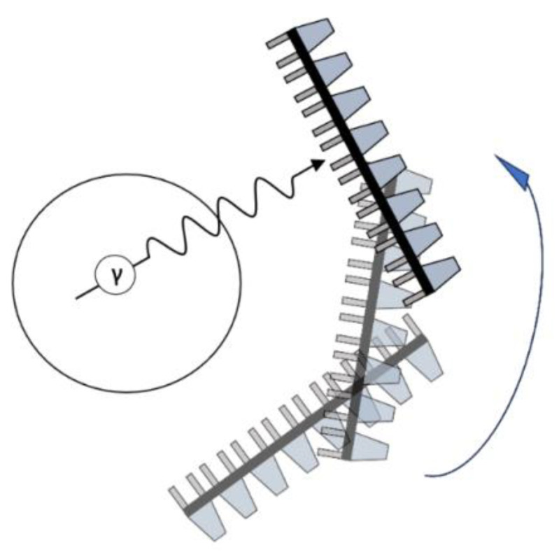
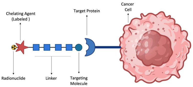
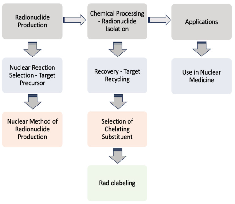
Scandium (Sc) isotopes have recently attracted significant attention in the search for new radionuclides with potential uses in personalized medicine, especially in the treatment of specific cancer patient categories. In particular, Sc-43 and Sc-44, as positron emitters with a satisfactory half-life (3.9 and 4.0 h, respectively), are ideal for cancer diagnosis via Positron Emission Tomography (PET). On the other hand, Sc-47, as an emitter of beta particles and low gamma radiation, may be used as a therapeutic radionuclide, which also allows Single-Photon Emission Computed Tomography (SPECT) imaging. As these scandium isotopes follow the same biological pathway and chemical reactivity, they appear to fit perfectly into the "theranostic pair" concept. A step-by-step description, initiating from the moment of scandium isotope production and leading up to their preclinical and clinical trial applications, is presented. Recent developments related to the nuclear reactions selected and employed to produce the radionuclides Sc-43, Sc-44, and Sc-47, the chemical processing of these isotopes and the main target recovery methods are also included. Furthermore, the radiolabeling of the leading chelator, 1,4,7,10-tetraazacyclododecane-1,4,7,10-tetraacetic acid (DOTA), and its structural analogues with scandium is also discussed and the advantages and disadvantages of scandium complexation are evaluated. Finally, a review of the preclinical studies and clinical trials involving scandium, as well as future challenges for its clinical uses and applications, are presented.
Keywords: DOTA; chelating agents; nuclear medicine; scandium imaging and diagnostic agents; scandium radiolabeled complexes production and development; scandium radiopharmaceuticals; scandium theranostic pair.
Conflict of interest statement
The authors declare no conflicts of interest.
Figures

Similar articles
-
Promising Scandium Radionuclides for Nuclear Medicine: A Review on the Production and Chemistry up to In Vivo Proofs of Concept.Cancer Biother Radiopharm. 2018 Oct;33(8):316-329. doi: 10.1089/cbr.2018.2485. Epub 2018 Sep 28. Cancer Biother Radiopharm. 2018. PMID: 30265573 Review.
-
Scandium and terbium radionuclides for radiotheranostics: current state of development towards clinical application.Br J Radiol. 2018 Nov;91(1091):20180074. doi: 10.1259/bjr.20180074. Epub 2018 Jun 15. Br J Radiol. 2018. PMID: 29658792 Free PMC article. Review.
-
Optimization of reaction conditions for the radiolabeling of DOTA and DOTA-peptide with (44m/44)Sc and experimental evidence of the feasibility of an in vivo PET generator.Nucl Med Biol. 2014 May;41 Suppl:e36-43. doi: 10.1016/j.nucmedbio.2013.11.004. Epub 2013 Nov 16. Nucl Med Biol. 2014. PMID: 24361353
-
Macrocyclic complexes of scandium radionuclides as precursors for diagnostic and therapeutic radiopharmaceuticals.J Inorg Biochem. 2011 Feb;105(2):313-20. doi: 10.1016/j.jinorgbio.2010.11.003. Epub 2010 Nov 13. J Inorg Biochem. 2011. PMID: 21194633
-
Promises of cyclotron-produced 44Sc as a diagnostic match for trivalent β--emitters: in vitro and in vivo study of a 44Sc-DOTA-folate conjugate.J Nucl Med. 2013 Dec;54(12):2168-74. doi: 10.2967/jnumed.113.123810. Epub 2013 Nov 6. J Nucl Med. 2013. PMID: 24198390
Cited by
-
Impact of Dietary Salicylates on Iron, Zinc, and Copper Status in Preeclampsia Model Rats Induced by L-NAME.Biol Trace Elem Res. 2025 Aug 7. doi: 10.1007/s12011-025-04772-1. Online ahead of print. Biol Trace Elem Res. 2025. PMID: 40773053
-
Synthesis of DOTA-Based 43Sc Radiopharmaceuticals Using Cyclotron-Produced 43Sc as Exemplified by [43Sc]Sc-PSMA-617 for PSMA PET Imaging.Methods Protoc. 2025 Jun 4;8(3):58. doi: 10.3390/mps8030058. Methods Protoc. 2025. PMID: 40559446 Free PMC article.
References
-
- Jalilian A.R., Gizawy M.A., Alliot C., Takacs S., Chakarborty S., Rovais M.R.A., Pupillo G., Nagatsu K., Park J.H., Khandaker M.U., et al. IAEA activities on 67Cu, 186Re, 47Sc theranostic radionuclides and radiopharmaceuticals. Curr. Radiopharm. 2021;14:306–314. doi: 10.2174/1874471013999200928162322. - DOI - PubMed
-
- Srivastava S.C., Mausner L.F. Therapeutic Nuclear Medicine. Springer; Berlin/Heidelberg, Germany: 2013. Therapeutic radionuclides: Production, physical characteristics, and applications; pp. 11–50.
-
- Payolla F.B., Massabni A.C., Orvig C. Radiopharmaceuticals for diagnosis in nuclear medicine: A short review. Eclética Química. 2019;44:11–19.
-
- Ilem-Ozdemir D., Gundogdu E.A., Ekinci M., Ozgenc E., Asikoglu M. Biomedical Applications of Nanoparticles. William Andrew Publishing; Norwich, NY, USA: 2019. Nuclear medicine and radiopharmaceuticals for molecular diagnosis; pp. 457–490.
Publication types
MeSH terms
Substances
LinkOut - more resources
Full Text Sources
Research Materials
