Review
. 2024 May 30;25(11):6003.
doi: 10.3390/ijms25116003.

CAFs-Associated Genes (CAFGs) in Pancreatic Ductal Adenocarcinoma (PDAC) and Novel Therapeutic Strategy

Affiliations
Review

CAFs-Associated Genes (CAFGs) in Pancreatic Ductal Adenocarcinoma (PDAC) and Novel Therapeutic Strategy

Keishi Yamashita et al. Int J Mol Sci. .

Abstract

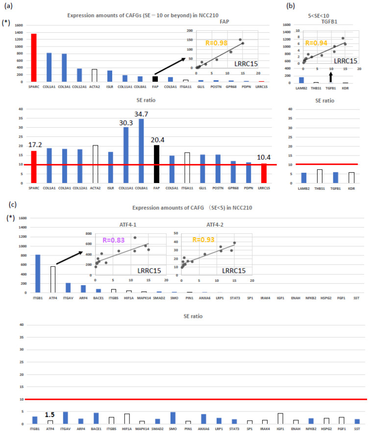
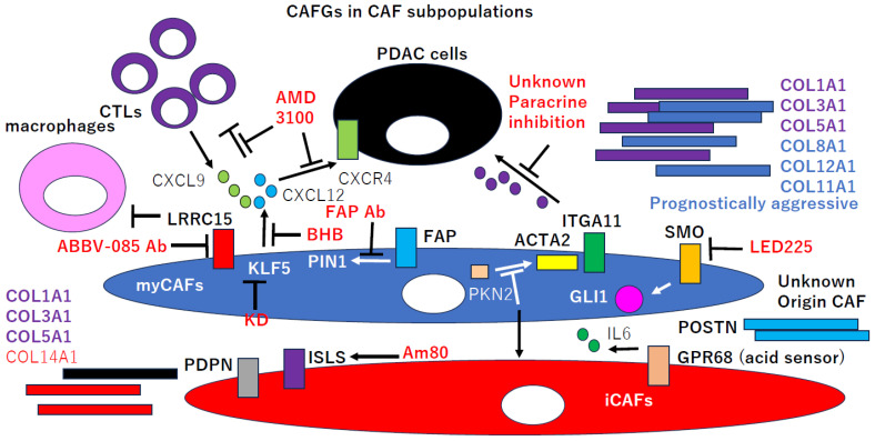
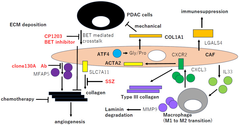
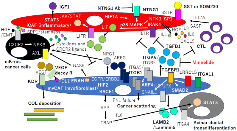
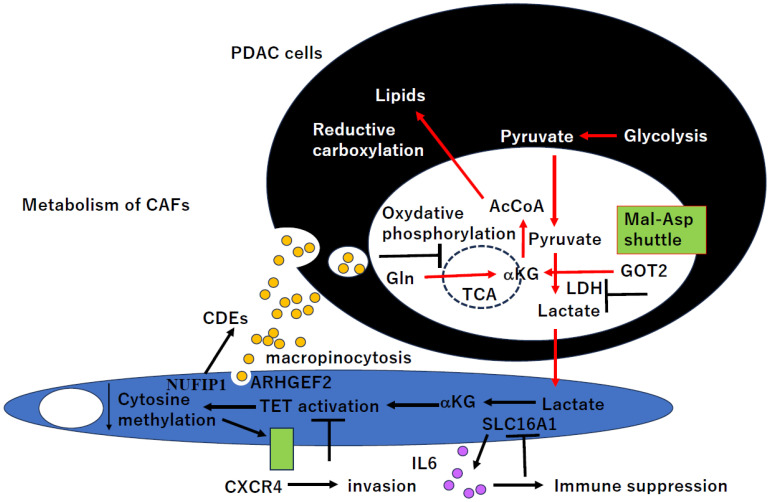
Pancreatic ductal adenocarcinoma (PDAC) is the most aggressive cancer with striking fibrosis, and its mortality rate is ranked second across human cancers. Cancer-associated fibroblasts (CAFs) play a critical role in PDAC progression, and we reviewed the molecular understanding of PDAC CAFs and novel therapeutic potential at present. CAFs-associated genes (CAFGs) were tentatively classified into three categories by stroma specificity representing stroma/epithelia expression ratios (SE ratios). The recent classification using single cell transcriptome technology clarified that CAFs were composed of myofibroblasts (myCAFs), inflammatory CAFs (iCAFs), and other minor ones (e.g., POSTN-CAFs and antigen presenting CAFs, apCAFs). LRRC15 is a myCAFs marker, and myCAFs depletion by diphtheria toxin induces the rapid accumulation of cytotoxic T lymphocytes (CTLs) and therefore augment PDL1 antibody treatments. This finding proposes that myCAFs may be a critical regulator of tumor immunity in terms of PDAC progression. myCAFs are located in CAFs adjacent to tumor cells, while iCAFs marked by PDPN and/or COL14A1 are distant from tumor cells, where hypoxic and acidic environments being located in iCAFs putatively due to poor blood supply is consistent with HIF1A and GPR68 expressions. iCAFs may be shared with SASP (secretion-associated phenotypes) in senescent CAFs. myCAFs are classically characterized by CAFGs induced by TGFB1, while chemoresistant CAFs with SASP may dependent on IL6 expression and accompanied by STAT3 activation. Recently, it was found that the unique metabolism of CAFs can be targeted to prevent PDAC progression, where PDAC cells utilize glucose, whereas CAFs in turn utilize lactate, which may be epigenetically regulated, mediated by its target genes including CXCR4. In summary, CAFs have unique molecular characteristics, which have been rigorously clarified as novel therapeutic targets of PDAC progression.

Keywords: CAFs; LRRC15; PDAC; SPARC; iCAFs; metabolism; myCAFs.


Conflict of interest statement

The authors declare no conflict of interest.

Figures

Figure 1
Figure 1
Molecular features of CAFGs, semi-CAFGs, and L-CAFGs in GSE35602. (a) Upper panel: Expression amounts of CAFGs (SE = 10 or beyond) in NCC210 (asterisks indicate expression amounts adjusted by GAPDHx100). Lower panel: SE ratio value is a y-axis. Inlet: FAP expression associated with LRRC15 expression in cancer stroma (R = 0.98). Red bars represent standard genes (SPARC and LRRC15), and black bars are CAFs markers (FAP in the figures). (b) Upper panel: Expression amounts of CAFGs (5 < SE < 10) in NCC210. Lower panel: SE ratio. Inlet: TGFB1 expression associated with LRRC15 expression in cancer stroma (R = 0.94). (c) Upper panel: Expression amounts of CAFGs (SE < 5) in NCC210. Lower panel: SE ratio. Inlet: ATF4 (probe 1 and probe 2) expression associated with LRRC15 expression in cancer stroma (R = 0.83 and 0.93, respectively).
Figure 2
Figure 2
CAFGs in PDAC and novel therapeutic potential. Therapeutic potential of targets of CAFGs (LRCC15, FAP, ACTA2, ITGA11, and GLI1) on myCAFs and those (PDPN, ISLS, and GPR68) on iCAFs. CAFGs also include collagen family genes such as myCAFs collagen (COL8A1, COL11A1, and COL12A1), iCAF collagen (COL14A1), and panCAF collagen (COL1A1, COL3A1, and COL5A1). POSTN is an ECM ascribed to CAFGs with unknown origin.
Figure 3
Figure 3
CAFGs collagens in PDAC and novel therapeutic potential. Therapeutic potential of targets of collagen production and deposits by MFAP5, BET, and SLC11A7. CAFGs (LRCC15, FAP, ACTA2, ITGA11, and GLI1) in myCAFs and those (PDPN, ISLS, and GPR68) in iCAFs. COL1A1 is produced by ATF4, and Type III collagen is produced and mediated uniquely by CXC3/CXCR2 in PDAC. LGASL4 is an ECM involved in immune suppression in PDAC.
Figure 4
Figure 4
Semi-CAFGs and L-CAFGs in PDAC and their novel therapeutic potential. TGFB1 is involved in myCAFs activation and iCAFs suppression and is suppressed by Minnelide. TGFB1 induces other CAFGs such as LRRC15 and ITGA11 as well as SNAIL1. For CAFs activation, ITGA5/ITGB1 internalization by ARF4 is essential and mediated by ITGAV/ITGB5. CAFs-secreted LAMB2 (Laminn5) may be critical for acinar-ductal transdifferentiation and STAT3 activation and is mediated by ITGA4. KDR (VEGFR) could be targeted for collagen deposition control. HIF1A is critical for iCAFs activation, while HIF2 is important for myCAFs activation. myCAFs subpopulations utilize EGFR/ERBB2 activation by AREG and/or NRG1, while iCAFs use p38 MAPK (MAPK14)/NFKB activation pathway to induce SASP fibroblast phenotypes.
Figure 5
Figure 5
CAFs metabolism in PDAC and novel therapeutic potential. PDAC cells suppressed oxidative phosphorylation (TCA cycle), while glycolysis to generate lactate and reductive carboxylation are uniquely activated and mediated by CAFs-derived exosomes (CDEs) in PDAC cells. Lactate from PDAC cells were utilized by CAFs, and CXCR4 expression is epigenetically affected by this unique metabolism of CAFs.

Similar articles

Cited by

References

    1. Rahib L., Smith B.D., Aizenberg R., Rosenzweig A.B., Fleshman J.M., Matrisian L.M. Projecting cancer incidence and deaths to 2030: The unexpected burden of thyroid, liver, and pancreas cancers in the United States. Cancer Res. 2014;74:2913–2921. doi: 10.1158/0008-5472.can-14-0155. - DOI - PubMed
    1. Raphael B.J., Hruban R.H., Aguirre A.J., Moffitt R.A., Yeh J.J., Stewart C., Lolla L. Integrated Genomic Characterization of Pancreatic Ductal Adenocarcinoma. Cancer Cell. 2017;32:185–203.e113. doi: 10.1016/j.ccell.2017.07.007. - DOI - PMC - PubMed
    1. Shaashua L., Ben-Shmuel A., Pevsner-Fischer M., Friedman G., Levi-Galibov O., Nandakumar S., Barki D., Nevo R., Brown L.E., Zhang W., et al. BRCA mutational status shapes the stromal microenvironment of pancreatic cancer linking clusterin expression in cancer associated fibroblasts with HSF1 signaling. Nat. Commun. 2022;13:6513. doi: 10.1038/s41467-022-34081-3. - DOI - PMC - PubMed
    1. Neuzillet C., Tijeras-Raballand A., Ragulan C., Cros J., Patil Y., Martinet M., Kocher H.M. Inter- and intra-tumoural heterogeneity in cancer-associated fibroblasts of human pancreatic ductal adenocarcinoma. J. Pathol. 2019;248:51–65. doi: 10.1002/path.5224. - DOI - PMC - PubMed
    1. Sahai E., Astsaturov I., Cukierman E., DeNardo D.G., Egeblad M., Evans R.M., Fearon D., Greten F.R., Hingorani S.R., Hunter T., et al. A framework for advancing our understanding of cancer-associated fibroblasts. Nat. Rev. Cancer. 2020;20:174–186. doi: 10.1038/s41568-019-0238-1. - DOI - PMC - PubMed

MeSH terms

Grants and funding