. 2024 May 31;25(11):6064.
doi: 10.3390/ijms25116064.

Characterization of Undiscovered miRNA Involved in Tumor Necrosis Factor Alpha-Induced Atrophy in Mouse Skeletal Muscle Cell Line

Affiliations

Characterization of Undiscovered miRNA Involved in Tumor Necrosis Factor Alpha-Induced Atrophy in Mouse Skeletal Muscle Cell Line

Dominika Pigoń-Zając et al. Int J Mol Sci. .

Abstract

Muscular atrophy is a complex catabolic condition that develops due to several inflammatory-related disorders, resulting in muscle loss. Tumor necrosis factor alpha (TNF-α) is believed to be one of the leading factors that drive inflammatory response and its progression. Until now, the link between inflammation and muscle wasting has been thoroughly investigated, and the non-coding RNA machinery is a potential connection between the candidates. This study aimed to identify specific miRNAs for muscular atrophy induced by TNF-α in the C2C12 murine myotube model. The difference in expression of fourteen known miRNAs and two newly identified miRNAs was recorded by next-generation sequencing between normal muscle cells and treated myotubes. After validation, we confirmed the difference in the expression of one novel murine miRNA (nov-mmu-miRNA-1) under different TNF-α-inducing conditions. Functional bioinformatic analyses of nov-mmu-miRNA-1 revealed the potential association with inflammation and muscle atrophy. Our results suggest that nov-mmu-miRNA-1 may trigger inflammation and muscle wasting by the downregulation of LIN28A/B, an anti-inflammatory factor in the let-7 family. Therefore, TNF-α is involved in muscle atrophy through the modulation of the miRNA cellular machinery. Here, we describe for the first time and propose a mechanism for the newly discovered miRNA, nov-mmu-miRNA-1, which may regulate inflammation and promote muscle atrophy.

Keywords: TNF-α; inflammation; miRNA; muscle atrophy; myotubes.


Conflict of interest statement

The authors have no relevant financial or non-financial interests to disclose.

Figures

Figure 1
Figure 1
Model of C2C12 myotube atrophy. (A) Observable differences in myotube morphology under a microscope examination between untreated cells (well-differentiated myotubes) and C2C12 cell culture treated with 100 ng/mL TNF-α within 72 h (atrophy model). (B) Differences in the atrophy marker expressions—FBXO32 and MuRF1—between the control and TNF-α treated cells. (**—p < 0.001).
Figure 2
Figure 2
Identification of DEnov-miRNAs and DEmiRNAs between treated and control C2C12 cultures by next-generation sequencing (NGS) in a discovery stage. (A) Volcano plot illustrating differences in expression of 175 novel mmu-miRNAs between studied cultures—the significant difference in the expression of the two molecules was recorded for the samples. (B) Heat map of the two DEnov-miRNAs, demonstrating the considerable difference in expression among the tested samples. (C) Volcano plot illustrating the differences in the expression of 868 known mmu-miRNA sequences between the studied cultures—the significant difference is the expression of the top 14 molecules. (D) Heat map illustrating expression differences of the top 14 DEmiRNAs between treated C2C12 myotube cultures and control cells. Above all, 16 mmu-miRNAs were selected in the discovery stage: 6 miRNAs were upregulated and 10 downregulated.
Figure 3
Figure 3
Result of validation of the identified DEnov-miRNAs by qRT-PCR. (A) Differences in nov-mmu-miRNA-1 expression between the control and TNF-α-treated C2C12 cells at different time intervals (12, 24, 28, and 72 h, respectively) and differences in miRNA expression after 72 h of cell growth between the control and treated cells treated with different TNF-α concentrations (10, 50, 100, and 200 ng/mL, respectively). (B) Differences in nov-mmu-miRNA-2 expression between the control and TNF-α-treated C2C12 cells at different time intervals (12, 24, 28, and 72 h, respectively) and differences in the miRNA expression after 72 h of cell growth between the control and treated cells treated with different TNF-α concentrations (10, 50, 100, and 200 ng/mL, respectively) (* p < 0.05; ** p < 0.001; ns—non-significant).
Figure 4
Figure 4
Structural and functional analysis of nov-mmu-miRNA-1. (A) RNA secondary structure of nov-mmu-miRNA-1 (the mature sequence is highlighted in red, and the star sequence is in blue) and the result of GO enrichment analysis—the top 10 terms were presented for the biological process. (B) Cellular component. (C) Molecular function. (D) The result of KEGG enrichment analysis. (E) The top 10 enriched terms.
Figure 5
Figure 5
UpSet plot highlights several mutual targets for identified DEmiRNAs. (A) Horizontal bars represent several gene targets for the individual miRNAs, and the vertical bars demonstrate individual or mutual gene targets for intersection. Points and connecting lines in the matrix indicate sets that are part of the intersection. Red dots and the connecting lines demonstrate mutual gene targets between nov-mmu-miRNA-1 and at least two other selected DEmiRNAs. The three sets meeting these criteria were identified and summarized in the Venn diagrams (joint targets plot A—RAB11FIB4, plot B—RICTOR, and plot C—CNOT6L). (B) KEGG pathway enrichment analysis for the three genes targeted by nov-mmu-miRNA-1 and at least two other miRNAs. (C) Varied mice muscle tissue expression of RAB11FIP4, RICTOR, and CNOT6L—the columns in the chart represent gene expression in BM—biceps muscle, MT—muscle tissue, and SMT—skeletal muscle tissue. In contrast, the rows demonstrate their expression in the different study sets. The colors of cells refer to expression (white—no evidence, gray—very low, light blue—low, and deep blue—moderate).
Figure 6
Figure 6
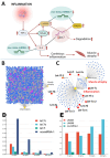
Potential link between nov-mmu-miRNA-1, muscle wasting, and its pro-inflammatory action. (A) Proposed mechanism of nov-mmu-miRNA-1 action leading to continuous inflammatory response and muscle wasting. (B) Molecular network of interactions between genes targeted by nov-mmu-miRNA-1 (purple dots), other miRNAs targeting those genes (blue squares), and inflammatory and atrophic conditions (yellow marks). (C) miRNAs extracted from the molecular network demonstrating selective involvement in the modulation of inflammatory and atrophic conditions (red squares represent let-7 family members indirectly regulated by nov-mmu-miRNA-1. (D) Significant expression changes of let-7c,e,i and nov-mmu-miRNA-1 under the different TNF-α concentrations. (E) Changes in LIN-28A and LIN-28B expression under the different TNF-α concentrations.

Similar articles

Cited by

References

    1. Wu Y., Che J., Jia P., Ma Y., Han Q., Wang X., Fu L., Dou H., Zheng Y. Near-infrared light-triggered polypyrrole promotes C2C12 cell differentiation and inhibits TNF-α induced myotube atrophy. Cell. Signal. 2022;100:110463. doi: 10.1016/j.cellsig.2022.110463. - DOI - PubMed
    1. Hitachi K., Honda M., Tsuchida K. The Functional Role of Long Non-Coding RNA in Myogenesis and Skeletal Muscle Atrophy. Cells. 2022;11:2291. doi: 10.3390/cells11152291. - DOI - PMC - PubMed
    1. Shen Y., Zhang R., Xu L., Wan Q., Zhu J., Gu J., Huang Z., Ma W., Shen M., Ding F., et al. Microarray Analysis of Gene Expression Provides New Insights Into Denervation-Induced Skeletal Muscle Atrophy. Front. Physiol. 2019;10:1298. doi: 10.3389/fphys.2019.01298. - DOI - PMC - PubMed
    1. Mukund K., Subramaniam S. Skeletal muscle: A review of molecular structure and function, in health and disease. Wiley Interdiscip. Rev. Syst. Biol. Med. 2020;12:e1462. doi: 10.1002/wsbm.1462. - DOI - PMC - PubMed
    1. LEE M.K., CHOI Y.H., NAM T.J. Pyropia yezoensis protein protects against TNF-α-induced myotube atrophy in C2C12 myotubes via the NF-κB signaling pathway. Mol. Med. Rep. 2021;24:486. doi: 10.3892/mmr.2021.12125. - DOI - PMC - PubMed

MeSH terms