

The Severity of Diabetic Retinopathy Corresponds with Corneal Nerve Alterations and Ocular Discomfort of the Patient
- PMID: 38892258
- PMCID: PMC11173272
- DOI: 10.3390/ijms25116072
The Severity of Diabetic Retinopathy Corresponds with Corneal Nerve Alterations and Ocular Discomfort of the Patient
Abstract
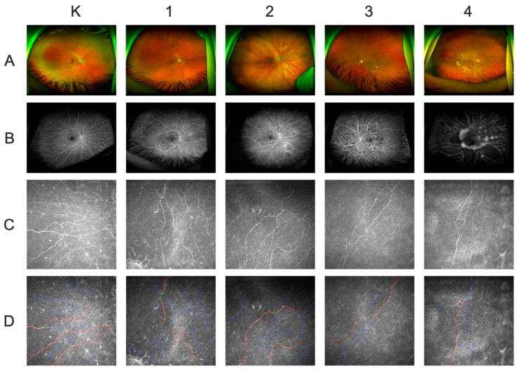
Diabetic retinopathy (DR) remains the leading cause of blindness in the working-age population. Its progression causes gradual damage to corneal nerves, resulting in decreased corneal sensitivity (CS) and disruption of anterior-eye-surface homeostasis, which is clinically manifested by increased ocular discomfort and dry eye disease (DED). This study included 52 DR patients and 52 sex- and age-matched controls. Ocular Surface Disease Index (OSDI) survey, tear film-related parameters, CS, and in vivo corneal confocal microscopy (IVCM) of the subbasal plexus were performed. Furthermore, all patients underwent tear sampling for neurotrophin and cytokine analysis. OSDI scores were greater in DR patients than in controls (p = 0.00020). No differences in the Schirmer test score, noninvasive tear film-break-up time (NIBUT), tear meniscus or interferometry values, bulbar redness, severity of blepharitis or meibomian gland loss were found. In the DR group, both the CS (p < 0.001), and the scotopic pupil diameter (p = 0.00008) decreased. IVCM revealed reduced corneal nerve parameters in DR patients. The stage of DR was positively correlated with the OSDI (Rs = +0.51, 95% CI: + 0.35-+0.64, p < 0.001) and negatively correlated with IVCM corneal nerve parameters and scotopic pupillometry (Rs = -0.26, 95% CI: -0.44--0.06, p = 0.0097). We found negative correlations between the OSDI and IVCM corneal innervation parameters. The DR group showed lower tear film-brain-derived neurotrophic factor (BDNF) levels (p = 0.0001) and no differences in nerve growth factor (NGF)-β, neurotrophin (NT)-4, vascular endothelial growth factor (VEGF), interleukin (IL)-1β, IL-4, IL-5, IL-6, or IL-12 concentrations. Tumor necrosis factor (TNF)-α, IL-2, IL-8, IL-10, granulocyte macrophage colony-stimulating factor (GM-CSF), and interferon (IFN)-γ levels were decreased among patients with DR. Corneal innervation defects have a direct impact on patients' subjective feelings. The evolution of DR appears to be associated with corneal nerve alterations, emphasizing the importance of IVCM.
Keywords: BDNF; OSDI; confocal microscopy; corneal innervation; corneal sensitivity; cytokines; diabetic retinopathy; neurotrophins; tear film; type 2 diabetes mellitus.
Conflict of interest statement
The authors declare no conflicts of interest.
Figures
Similar articles
-
Comparison of tear cytokines and neuropeptides, ocular surface parameters, and corneal nerve structure in patients with early-stage diabetes mellitus and control subjects.Int Ophthalmol. 2025 Mar 22;45(1):119. doi: 10.1007/s10792-025-03502-9. Int Ophthalmol. 2025. PMID: 40119961
-
Tear Fluid Progranulin as a Noninvasive Biomarker for the Monitoring of Corneal Innervation Changes in Patients With Type 2 Diabetes Mellitus.Transl Vis Sci Technol. 2024 Jul 1;13(7):9. doi: 10.1167/tvst.13.7.9. Transl Vis Sci Technol. 2024. PMID: 38984913 Free PMC article.
-
Multimodal Approach in Dry Eye Disease Combining In Vivo Confocal Microscopy and HLA-DR Expression.Transl Vis Sci Technol. 2024 Aug 1;13(8):39. doi: 10.1167/tvst.13.8.39. Transl Vis Sci Technol. 2024. PMID: 39177993 Free PMC article.
-
Tear biomarkers in dry eye disease: Progress in the last decade.Indian J Ophthalmol. 2023 Apr;71(4):1190-1202. doi: 10.4103/IJO.IJO_2981_22. Indian J Ophthalmol. 2023. PMID: 37026250 Free PMC article. Review.
-
A Review of Imaging Biomarkers of the Ocular Surface.Eye Contact Lens. 2020 Mar;46 Suppl 2(Suppl 2):S84-S105. doi: 10.1097/ICL.0000000000000684. Eye Contact Lens. 2020. PMID: 31833999 Free PMC article. Review.
Cited by
-
Systematic Inflammation and Oxidative Stress Elevation in Diabetic Retinopathy and Diabetic Patients with Macular Edema.Int J Mol Sci. 2025 Apr 17;26(8):3810. doi: 10.3390/ijms26083810. Int J Mol Sci. 2025. PMID: 40332476 Free PMC article.
-
Differential Ophthalmological Profile in Patients with Coronary Artery Disease Coexisting with Type 2 Diabetes Mellitus: Elevated Tear Cytokine Concentrations.J Clin Med. 2024 Aug 20;13(16):4906. doi: 10.3390/jcm13164906. J Clin Med. 2024. PMID: 39201047 Free PMC article.
-
Tear-fluid-derived biomarkers of ocular complications in diabetes: a systematic review and meta-analysis.BMC Med. 2025 Feb 12;23(1):84. doi: 10.1186/s12916-025-03855-z. BMC Med. 2025. PMID: 39939938 Free PMC article.
References
MeSH terms
Substances
LinkOut - more resources
Full Text Sources
Medical
Research Materials
