. 2024 May 31;25(11):6061.
doi: 10.3390/ijms25116061.

Effect of Hydroxyurea on Morphology, Proliferation, and Protein Expression on Taenia crassiceps WFU Strain

Affiliations

Effect of Hydroxyurea on Morphology, Proliferation, and Protein Expression on Taenia crassiceps WFU Strain

Diana G Rios-Valencia et al. Int J Mol Sci. .

Abstract

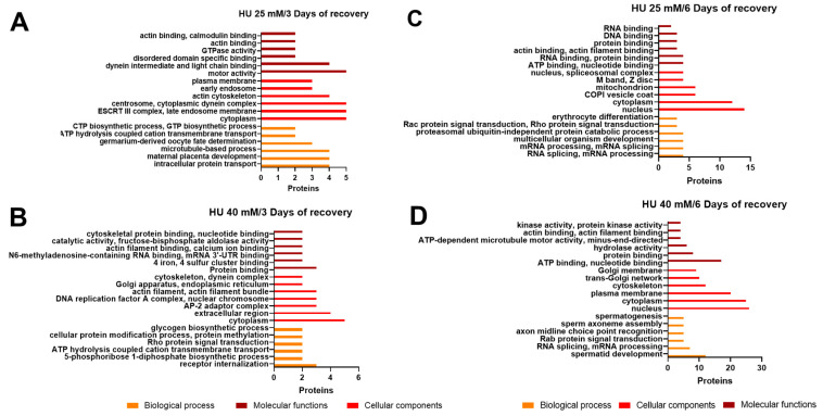
Flatworms are known for their remarkable regenerative ability, one which depends on totipotent cells known as germinative cells in cestodes. Depletion of germinative cells with hydroxyurea (HU) affects the regeneration of the parasite. Here, we studied the reduction and recovery of germinative cells in T. crassiceps cysticerci after HU treatment (25 mM and 40 mM of HU for 6 days) through in vitro assays. Viability and morphological changes were evaluated. The recovery of cysticerci's mobility and morphology was evaluated at 3 and 6 days, after 6 days of treatment. The number of proliferative cells was evaluated using EdU. Our results show morphological changes in the size, shape, and number of evaginated cysticerci at the 40 mM dose. The mobility of cysticerci was lower after 6 days of HU treatment at both concentrations. On days 3 and 6 of recovery after 25 mM of HU treatment, a partial recovery of the proliferative cells was observed. Proteomic and Gene Ontology analyses identified modifications in protein groups related to DNA binding, DNA damage, glycolytic enzymes, cytoskeleton, skeletal muscle, and RNA binding.

Keywords: Taenia crassiceps; cysticercus; hydroxyurea; proliferative cells; proteome.


Conflict of interest statement

The authors declare no conflicts of interest.

Figures

Figure 1
Figure 1
In vitro evaluation of the effect of hydroxyurea (HU) in T. crassiceps cysticerci. (A) Changes in the shape of cysticerci were detected by stereomicroscopy. Two concentrations of HU (25 mM and 40 mM) were applied for 6 days; the cysticerci were also evaluated after 3 and 6 days of recovery in HU-free medium. Black arrows show evaginated cysticerci. Scale bar represent 2 mm. (B) Changes in metabolic activity were measured through the reduction of Alamar Blue® (Y-axis) over a period of 24 h in the different groups (X-axis). **** p < 0.0001, *** p = 0.0001, and * p = 0.01.
Figure 2
Figure 2
The effect of HU treatment on the number of proliferative cells in the tissue of T. crassiceps cysticerci. (A) Whole-mount localization of proliferative cells (indicated with white arrows) on the bladder wall tissue of T. crassiceps WFU cysticerci. Subsequently, the mounts were stained with EdU (red) and DAPI (blue) to mark proliferative cells in phase S and cell nuclei, respectively. Bars represent 200 µm; n = 10–12 individual larvae in all groups. (B) The effect of HU treatment (25 mM or 40 mM) on the number of proliferative cells in the tissue from the cysticerci. Also, the effects of 3 and 6 days of recovery in in vitro cultures are shown. Groups of larvae without HU treatment were used as controls. The statistical analysis of the results was carried out using a two-way ANOVA and the Dunnet comparison test. **** p < 0.0001, *** p = 0.0001, ** p = 0.001, and * p = 0.01.
Figure 3
Figure 3
Proliferative cells in T. crassiceps WFU cysticerci after 6 days of HU treatment. Proliferative cells (phase S) (indicated with white arrows) were observed after staining with EdU (red); DAPI (blue) was used to counterstain cell nuclei. Here, representative pictures of whole mounts of (A) the bladder walls and (B) the scolices are shown. Bars represent 200 µm.
Figure 4
Figure 4
Proteins expressed after HU treatment. Venn diagram of differentially expressed proteins in different groups: (A) 6 days of HU treatment, (B) 3 days of recovery, and (C) 6 days of recovery. Only proteins with annotation are present in the figure.
Figure 5
Figure 5
Volcano plot of differentially expressed proteins identified through mass spectrometry. The X-axis represents the log2 of expression fold change whereas the Y-axis represents -Log10 negative logarithm base 10 (-Log10) of the p-value. In green are indicating statistical significance values where a p-value ≤ 0.05. Downregulated proteins are shown on the right side of the plot (HU high/control low), whereas upregulated proteins are on the left side (HU low/control high). Non-significant changes are displayed in grey. (A) Control vs. HU, 25 mM, and 6 days of treatment; (B) Control vs. HU, 40 mM, and 6 days of treatment; (C) Control vs. HU, 25 mM, and 3 days of recovery; (D) Control vs. HU, 40 mM, and 3 days of recovery; (E) Control vs. HU, 25 mM, and 6 days of recovery; and (F) Control vs. HU, 40 mM, and 6 days of recovery.
Figure 6
Figure 6
Gene ontology (GO) analysis of differentially expressed proteins after HU treatment at concentrations of (A) 25 mM and (B) 40 mM. Within the three ontologies, Biological Processes (BP): orange; Cellular Components (CC): red; Molecular Function (MF): dark red; the top five protein classes are shown. All classes are included in Supplementary Table S3.
Figure 7
Figure 7
Gene ontology (GO) analyses of differentially expressed proteins after HU treatment and 3 days of recovery. Within the three ontologies (BP: orange; CC: red; and MF: dark red), the top five protein classes are shown. (A) HU 25 mM, 3 days of recovery; (B) HU 40 mM, 3 days of recovery; (C) HU 25 mM, 6 days of recovery; and (D) HU 40 mM, 6 days of recovery. All classes are included in Supplementary Table S3.

Similar articles

References

    1. Loos-Frank B. An up-date of Verster’s (1969) ‘Taxonomic revision of the genus Taenia Linnaeus’ (Cestoda) in table format. Syst. Parasitol. 2000;45:155–183. doi: 10.1023/A:1006219625792. - DOI - PubMed
    1. Freeman R.S., Fallis A.M., Shea M., Maberley A.L., Walters J. Intraocular Taenia crassiceps (Cestoda). II. The parasite. Am. J. Trop. Med. Hyg. 1973;22:493–495. doi: 10.4269/ajtmh.1973.22.493. - DOI - PubMed
    1. Heldwein K., Biedermann H.G., Hamperl W.D., Bretzel G., Löscher T., Laregina D., Frosch M., Büttner D.W., Tappe D. Subcutaneous Taenia crassiceps infection in a patient with non-Hodgkin’s lymphoma. Am. J. Trop. Med. Hyg. 2006;75:108–111. doi: 10.4269/ajtmh.2006.75.108. - DOI - PubMed
    1. Goesseringer N., Lindenblatt N., Mihic-Probst D., Grimm F., Giovanoli P. Taenia crassiceps upper limb fasciitis in a patient with untreated acquired immunodeficiency syndrome and chronic hepatitis C infection—The role of surgical debridement. J. Plast. Reconstr. Aesthet. Surg. 2011;64:e174–e176. doi: 10.1016/j.bjps.2011.02.011. - DOI - PubMed
    1. Ntoukas V., Tappe D., Pfütze D., Simon M., Holzmann T. Cerebellar cysticercosis caused by larval Taenia crassiceps tapeworm in immunocompetent woman, Germany. Emerg. Infect. Dis. 2013;19:2008–2011. doi: 10.3201/eid1912.130284. - DOI - PMC - PubMed

LinkOut - more resources