. 2024 May 31;25(11):6089.
doi: 10.3390/ijms25116089.

Cell Wall Microdomains in the External Glands of Utricularia dichotoma Traps

Affiliations

Cell Wall Microdomains in the External Glands of Utricularia dichotoma Traps

Bartosz J Płachno et al. Int J Mol Sci. .

Abstract

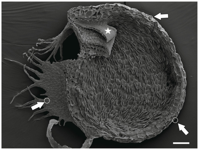
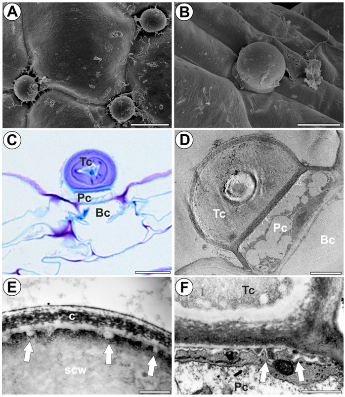
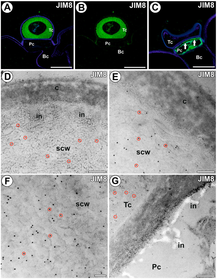
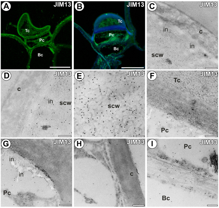
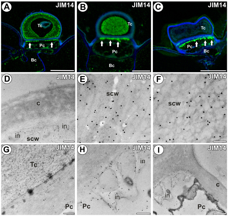
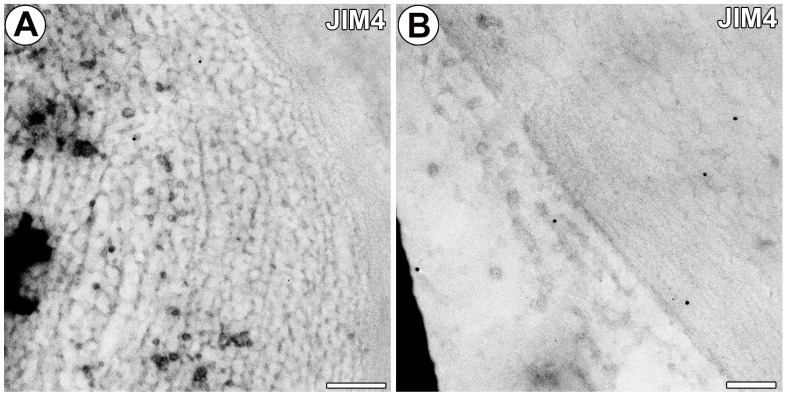
The genus Utricularia (bladderworts) species are carnivorous plants that prey on invertebrates using traps with a high-speed suction mechanism. The outer trap surface is lined by dome-shaped glands responsible for secreting water in active traps. In terminal cells of these glands, the outer wall is differentiated into several layers, and even cell wall ingrowths are covered by new cell wall layers. Due to changes in the cell wall, these glands are excellent models for studying the specialization of cell walls (microdomains). The main aim of this study was to check if different cell wall layers have a different composition. Antibodies against arabinogalactan proteins (AGPs) were used, including JIM8, JIM13, JIM14, MAC207, and JIM4. The localization of the examined compounds was determined using immunohistochemistry techniques and immunogold labeling. Differences in composition were found between the primary cell wall and the cell secondary wall in terminal gland cells. The outermost layer of the cell wall of the terminal cell, which was cuticularized, was devoid of AGPs (JIM8, JIM14). In contrast, the secondary cell wall in terminal cells was rich in AGPs. AGPs localized with the JIM13, JIM8, and JIM14 epitopes occurred in wall ingrowths of pedestal cells. Our research supports the hypothesis of water secretion by the external glands.

Keywords: Lentibulariaceae; arabinogalactan proteins; bladderworts; carnivorous plants; cell wall; cell wall microdomains; cuticle; glands; scanning transmission electron microscopy; transfer cells; trichomes.


Conflict of interest statement

The authors declare no conflicts of interest.

Figures

Figure 1
Figure 1
A sagittally halved trap of Utricularia dichotoma subsp. novae-zelandiae showing the external glands (arrow and white circle) and trap door (star), SEM, scale bar 200 µm.
Figure 2
Figure 2
Distribution, morphology, and structure of the external trap gland of Utricularia dichotoma subsp. novae-zelandiae. (A,B) Morphology of the external trap glands, bar 25 µm, and bar 10 µm. (C) Structure of external gland: terminal cell (Tc), pedestal cell (Pc), basal cell (Bc), bar 10 µm. (D) Ultrastructure of external gland: terminal cell (Tc), pedestal cell (Pc), basal cell (Bc), bar 3 µm. (E) Ultrastructure of the terminal cell, note outermost cuticularized layer of the cell wall (c), cell wall ingrowths (arrow), secondary cell wall (scw), bar 1 µm. (F) Ultrastructure of the terminal cell (Tc) and pedestal cell (Pc), cell wall ingrowths (arrow), bar 1 µm.
Figure 3
Figure 3
The arabinogalactan proteins detected in the external gland labeled with JIM8 (green color—signal of antibody, red circles—gold particle designation). (A,B) Arabinogalactan proteins (labeled with JIM8) were detected in the external gland; note strong fluorescence signal in the terminal cell; terminal cell (Tc), pedestal cell (Pc), basal cell (Bc), bar 10 µm and bar 10 µm. (C) Arabinogalactan proteins (labeled with JIM8) were detected in the external gland; note strong fluorescence signal in wall ingrowths in the pedestal cell (arrow); terminal cell (Tc), pedestal cell (Pc), basal cell (Bc), bar 10 µm. (DF) Immunogold labeling with JIM8 in the terminal cell; the outermost cuticularized layer of the cell wall (c), secondary cell wall (scw), wall ingrowths (in), bar 400 nm, bar 200 nm, bar 100 nm. (G) Immunogold labeling with JIM8 in the terminal cell (Tc) and pedestal cell (Pc), cell wall ingrowths (in), bar 400 nm.
Figure 4
Figure 4
The arabinogalactan proteins detected in the external gland labeled with JIM13 (green color—signal of antibody). (A,B) Arabinogalactan proteins (labeled with JIM13) were detected in the external gland; note strong fluorescence signal in the terminal cell; terminal cell (Tc), pedestal cell (Pc), basal cell (Bc), bar 10 µm and bar 10 µm. (CE) Immunogold labeling with JIM13 in the terminal cell; the outermost cuticularized layer of the cell wall (c), secondary cell wall (scw), wall ingrowths (in), bar 400 nm, bar 200 nm, bar 100 nm. (F) Immunogold labeling with JIM13 in the terminal cell (Tc) and pedestal cell (Pc), bar 300 nm. (G,H) Immunogold labeling with JIM13 in the pedestal cell (Pc), cell wall ingrowths (in), cuticularized later cell wall (c), bar 300 nm and bar 300 nm. (I) Immunogold labeling with JIM13 in the cell walls between pedestal cell and basal cell, bar 200 nm.
Figure 5
Figure 5
The arabinogalactan proteins detected in the external gland labeled with JIM14 (green color—signal of antibody). (A,B) Arabinogalactan proteins (labeled with JIM14) were detected in the external gland; note strong fluorescence signal in terminal cell; terminal cell (Tc), pedestal cell (Pc), basal cell (Bc), wall ingrowths in pedestal cell (arrow), bar 10 µm and bar 5 µm. (C) Arabinogalactan proteins (labeled with JIM14) were detected in the external gland; note strong fluorescence signal in wall ingrowths in pedestal cell (arrow); terminal cell (Tc), pedestal cell (Pc), basal cell (Bc), bar 10 µm. (DF) Immunogold labeling with JIM14 in the terminal cell; the outermost cuticularized layer of cell wall (c), secondary cell wall (scw), wall ingrowths (in), bar 200 nm, bar 100 nm, bar 100 nm. (G) Immunogold labeling with JIM14 in the terminal cell (Tc) and pedestal cell (Pc), bar 200 nm. (H,I) Immunogold labeling with JIM14 in the pedestal cell (Pc), cell wall ingrowths (in), cuticularized later cell wall (c), bar 200 nm, and bar 400 nm.
Figure 6
Figure 6
Immunogold labeling with JIM4 in the external gland. (A,B) Immunogold labeling with JIM4 in the terminal cell; note only a few gold particles, bar 200 nm and bar 100 nm.

Similar articles

Cited by

References

    1. Müller K., Borsch T., Legendre L., Porembski S., Theisen I., Barthlott W. Evolution of carnivory in Lentibulariaceae and the Lamiales. Plant Biol. 2004;6:477–490. doi: 10.1055/s-2004-817909. - DOI - PubMed
    1. Müller K.F., Borsch T., Legendre L., Porembski S., Barthlott W. Recent progress in understanding the evolution of carnivorous Lentibulariaceae (Lamiales) Plant Biol. 2006;8:748–757. doi: 10.1055/s-2006-924706. - DOI - PubMed
    1. Fonseca L.H.M. Combining molecular and geographical data to infer the phylogeny of Lamiales and its dispersal patterns in and out of the tropics. Mol. Phylogenetics Evol. 2021;164:107287. doi: 10.1016/j.ympev.2021.107287. - DOI - PubMed
    1. Lloyd F.E. The Carnivorous Plants. Chronica Botanica Company; Waltham, MA, USA: 1942.
    1. Heslop-Harrison Y. Scanning electron microscopy of fresh leaves of Pinguicula. Science. 1970;167:172–174. doi: 10.1126/science.167.3915.172. - DOI - PubMed

LinkOut - more resources