. 2024 May 31;25(11):6086.
doi: 10.3390/ijms25116086.

Secukinumab and Dead Sea Climatotherapy Impact Resolved Psoriasis Skin Differently Potentially Affecting Disease Memory

Affiliations

Secukinumab and Dead Sea Climatotherapy Impact Resolved Psoriasis Skin Differently Potentially Affecting Disease Memory

Thomas Emmanuel et al. Int J Mol Sci. .

Abstract

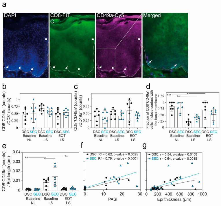
Secukinumab and Dead Sea treatment result in clear skin for many psoriasis patients, through distinct mechanisms. However, recurrence in the same areas after treatments suggests the existence of a molecular scar. We aimed to compare the molecular and genetic differences in psoriasis patients who achieved complete response from secukinumab and Dead Sea climatotherapy treatments. We performed quantitative immunohistochemical and transcriptomic analysis, in addition to digital spatial profiling of skin punch biopsies. Histologically, both treatments resulted in a normalization of the lesional skin to a level resembling nonlesional skin. Interestingly, the transcriptome was not normalized by either treatments. We revealed 479 differentially expressed genes between secukinumab and Dead Sea climatotherapy at the end of treatment, with a psoriasis panel identifying SERPINB4, SERPINB13, IL36G, IL36RN, and AKR1B10 as upregulated in Dead Sea climatotherapy compared with secukinumab. Using digital spatial profiling, pan-RAS was observed to be differentially expressed in the microenvironment surrounding CD103+ cells, and IDO1 was differentially expressed in the dermis when comparing the two treatments. The differences observed between secukinumab and Dead Sea climatotherapy suggest the presence of a molecular scar, which may stem from mechanistically different pathways and potentially contribute to disease recurrence. This may be important for determining treatment response duration and disease memory.

Keywords: T lymphocytes; biologics; inflammatory skin diseases; phototherapy; psoriasis; tissue resident memory T-cells.


Conflict of interest statement

T.E. reports being an investigator on investigator-initiated clinical trials supported by LEO Pharma and Janssen and has received honoraria as a speaker for Bristol Myers Squibb. T.L. is funded by LEO Pharma. L.I. has served as a consultant and/or paid speaker for and/or participated in clinical trials sponsored by the following: AbbVie, Almirall, Amgen, Astra Zeneca, Bristol Myers Squibb, Boehringer Ingelheim, Celgene, Centocor, Eli Lilly, Janssen-Cilag, Kyowa, LEO Pharma, MSD, Novartis, Pfizer, Regranion, Samsung, and UCB. Furthermore, L.I. is an employee at MC2 Therapeutics. C.J. has served as a consultant and/or paid speaker for Eli Lilly, LEO Pharma, AbbVie, and L’Oréal. With no relation to the present manuscript, T.B. has received research funding or educational grants from Novartis, AbbVie, and UCB, and honoraria as consultant and/or speaker from Eli Lilly, Novartis, Leo Pharma, and UCB.

Figures

Figure 1
Figure 1
Immunohistochemical results from the two cohorts at baseline and end of treatment. (a) Study schematic of the treatment duration from baseline to end of treatment (EOT) for patients treated with Dead Sea climatotherapy (DSC) and patients treated with secukinumab (SEC). (bf) Results from hematoxylin and eosin (HE) staining used to quantify the epidermal thickness and immunohistochemistry of Ki67+, CD3+, CD4+, and CD8+ cells from nonlesional (NL) and lesional (LS) skin at baseline, and LS skin at EOT. The dashed lines indicate the interface between the epidermis and dermis. Sequential slides from the same patient are shown. Scale bars = 200 μm. Mean ± SD depicted. One-way analysis of variance (ANOVA) with Tukey’s multiple comparison test was used to compare across cohorts, and repeated measures analysis of variance with post hoc Šidák test was used to compare between timepoints within studies. * p < 0.05, ** p < 0.01, *** p < 0.001.
Figure 2
Figure 2
Immunohistochemical results from CD15, CD56, CD103, CD207, CD163, CD207, and FOXP3 from the two cohorts at baseline and end of treatment. (ac) Results from quantitative immunohistochemistry analysis of CD15+, CD56+, CD103+, CD163+, CD207+, and FOXP3+ cells from nonlesional (NL) and lesional (LS) skin taken at baseline and end of treatment (EOT) from patients treated with Dead Sea climatotherapy (DSC) and patients treated with secukinumab (SEC). Black arrows show examples of CD103+ cells colocalizing with CD207+ cells. Scale bars = 200 μm. Mean ± SD depicted. One-way analysis of variance (ANOVA) with Tukey’s multiple comparison test was used to compare across cohorts, and repeated measures analysis of variance with post hoc Šidák test was used to compare between timepoints within studies. * p < 0.05, ** p < 0.01, *** p < 0.001.
Figure 3
Figure 3
Immunohistochemical results from CD8+ and CD49a+ staining. (a) Representative CD8 and CD49a staining from lesional skin (LS) showing the typical localization of tissue-resident memory T-cells near the epidermal dermal border. White arrows indicate examples of CD8+CD49a+ cells. The dashed line indicates the interface between epidermis and dermis. Scale bar = 100 μm. (bd) Results from quantitative immunohistochemistry analysis of CD8+/−CD49a+/− cells in the epidermis from baseline nonlesional (NL), LS, and end of treatment (EOT) LS skin. (e) CD8+CD49a+ cell counts normalized to epidermal length from DSC and SEC treatments at baseline and EOT. (f) Spearman correlation for PASI and CD8+CD49a+ cell counts for DSC and SEC treatments. (g) Pearson correlation for epidermal thickness and CD8+CD49a+ cell counts for DSC and SEC treatments. For figure (be), one-way analysis of variance (ANOVA) with Tukey’s multiple comparison test was used to compare across cohorts, and a mixed model analysis with post hoc Šidák test was used to compare between timepoints within studies. Mean ± SD depicted. * p < 0.05, ** p < 0.01, *** p < 0.001.
Figure 4
Figure 4
Microarray analysis of resolved skin from the two treatment cohorts. (a) Heatmap and two-way semi-supervised clustering based on 1217 DEGs between the SEC and DSC groups at end of treatment (Var > 0.1, p < 0.05, q = 0.10). The samples are colored according to study: DSC, black; SEC, cyan. The colors in the heatmap signify high (yellow) or low (blue) expression of a particular gene across samples (z-scaled values). (c) Heatmap and two-way semi-supervised clustering based on 48 psoriasis-defining genes. The samples are colored according to study: DSC, black; SEC, cyan. The colors in the heatmap signify high (yellow) or low (blue) expression of a particular gene across samples (z-scaled values). (b) Volcano plot showing the log2 (fold change) between the SEC and DSC groups at end of treatment for all genes on the x-axis and the −log10 of the p-value for the two groups’ (SEC vs. DSC) t-test on the y-axis. The genes that met the cut-off criteria (p < 0.05 and >2-fold change) are colored in the same way as for the heatmap (higher/lower in SEC, yellow/blue). (d) Heatmap and semi-supervised hierarchical clustering showing the six most significantly different upregulated and downregulated DEGs based on the 48 psoriasis-related genes.
Figure 5
Figure 5
Schematic of the digital spatial profiler workflow and assessment of CD45+ cells, MelanA+ cells, and the microenvironment in the skin. (a) (1) Slides were incubated with a cocktail of primary labeled fluorescent antibodies mixed with a panel of commercial NanoString antibodies linked to photocleavable barcoded oligos. (2) Areas of illumination (AOIs) were selected in the digital spatial profiler (DSP) using fluorescent morphology markers to distinguish tissues and cells. (3) Ultraviolet light separated the oligos from the antibodies. The oligos were then pulled by a microcapillary tip and deposited in individual wells on a 96-well plate. (4) Cells were manually pooled from several wells into one well and hybridized according to the NanoString protocol. (5) Oligos were counted in the nCounter and the results could then be imported into the DSP, allowing real-time spatial analysis of individual AOIs and normalizations of values. Real-time spatial analysis was limited in this experiment, due to the pooling of AOIs. (b) Image showing the setup on the slides with normal skin (NS), baseline nonlesional (NL), baseline lesional (LS), and EOT LS. Size bar = 1 mm. (c) Larger image of baseline LS showing the ROIs selected. Three 50 μm diameter ROIs from the dermis and epidermis were collected from a random area without visible CD3+, CD45+, or MelanA+ cells. Ten 10 μm diameter ROIs from CD45+ cells (yellow) and MelanA+ cells (blue) were collected from the epidermis and ten 10–20 μm width contour sections surrounding the CD45+ cells (CD45+ cell microenvironment) and MelanA+ cells (MelanA+ cell microenvironment) were selected using the polygon tool. Scale bar = 200 μm. (d) Heatmap and two-way unsupervised clustering based on the 70 most variable proteins (Var > 0.2) across samples. The samples are colored according to tissue (healthy skin, green; NL, brown; LS, gray) and histology. The colors in the heatmap signify high (yellow) or low (blue) expression of a particular gene across samples (z-scaled values). The samples clustered primarily according to dermis, but also according to CD45+ and MelanA+ cells. (e) PCA plot based on the same 70 proteins, colored according to histology.
Figure 6
Figure 6
Differentially expressed proteins at end of treatment between the two cohorts. (a) Differentially expressed proteins (DEPs) in the CD103+ microenvironment between Dead Sea climatotherapy (DSC) and secukinumab (SEC)-treated patients at end of treatment (EOT). (b) DEPs in the dermal infiltrate between DSC and SEC-treated patients at EOT. (c) DEPs in the epidermis between DSC- and SEC-treated patients at EOT.

References

    1. de Oliveira M.d.F.S.P., de Oliveira Rocha B., Duarte G.V. Psoriasis: Classical and emerging comorbidities. An. Bras. Dermatol. 2015;90:9–20. doi: 10.1590/abd1806-4841.20153038. - DOI - PMC - PubMed
    1. Griffiths C.E.M., Armstrong A.W., Gudjonsson J.E., Barker J.N.W.N. Psoriasis. Lancet. 2021;397:1301–1315. doi: 10.1016/S0140-6736(20)32549-6. - DOI - PubMed
    1. Parisi R., Iskandar I.Y.K.K., Kontopantelis E., Augustin M., Griffiths C.E.M.M., Ashcroft D.M. National, regional, and worldwide epidemiology of psoriasis: Systematic analysis and modelling study. BMJ. 2020;369:m1590. doi: 10.1136/bmj.m1590. - DOI - PMC - PubMed
    1. Reid C., Griffiths C. Psoriasis and Treatment: Past, Present and Future Aspects. Acta Derm. Venereol. 2020;100:70–80. doi: 10.2340/00015555-3386. - DOI - PMC - PubMed
    1. Mrowietz U., Steinz K., Gerdes S. Psoriasis: To treat or to manage? Exp. Dermatol. 2014;23:705–709. doi: 10.1111/exd.12437. - DOI - PubMed

Substances