. 2024 Jun 1;25(11):6102.
doi: 10.3390/ijms25116102.

Generation of iPSCs from a Patient with the M694V Mutation in the MEFV Gene Associated with Familial Mediterranean Fever and Their Differentiation into Macrophages

Affiliations

Generation of iPSCs from a Patient with the M694V Mutation in the MEFV Gene Associated with Familial Mediterranean Fever and Their Differentiation into Macrophages

Elena V Grigor'eva et al. Int J Mol Sci. .

Abstract

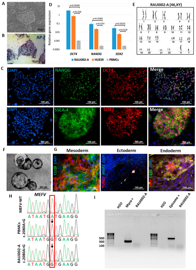
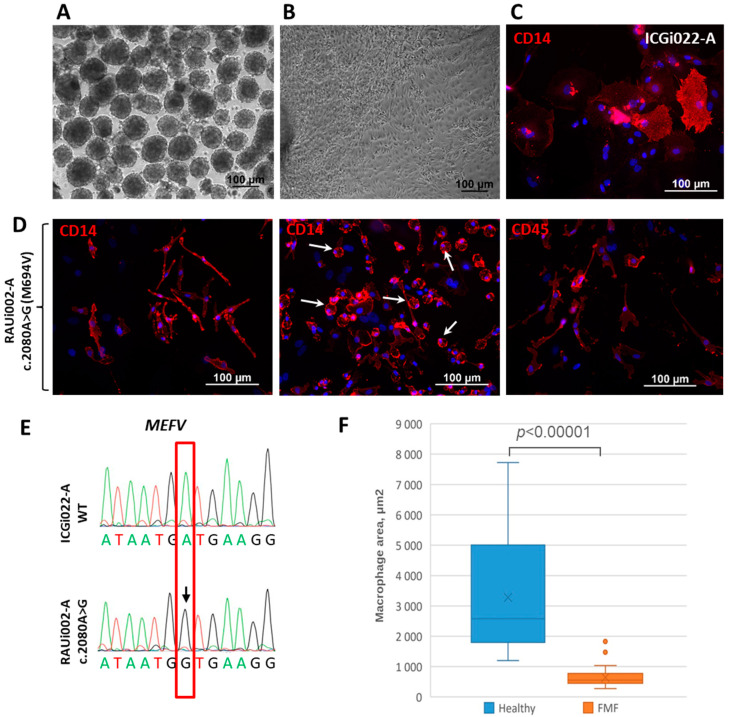
Familial Mediterranean fever (FMF) is a systemic autoinflammatory disorder caused by inherited mutations in the MEFV (Mediterranean FeVer) gene, located on chromosome 16 (16p13.3) and encoding the pyrin protein. Despite the existing data on MEFV mutations, the exact mechanism of their effect on the development of the pathological processes leading to the spontaneous and recurrent autoinflammatory attacks observed in FMF, remains unclear. Induced pluripotent stem cells (iPSCs) are considered an important tool to study the molecular genetic mechanisms of various diseases due to their ability to differentiate into any cell type, including macrophages, which contribute to the development of FMF. In this study, we developed iPSCs from an Armenian patient with FMF carrying the M694V, p.(Met694Val) (c.2080A>G, rs61752717) pathogenic mutation in exon 10 of the MEFV gene. As a result of direct differentiation, macrophages expressing CD14 and CD45 surface markers were obtained. We found that the morphology of macrophages derived from iPSCs of a patient with the MEFV mutation significantly differed from that of macrophages derived from iPSCs of a healthy donor carrying the wild-type MEFV gene.

Keywords: Familial Mediterranean fever; MEFV gene; differentiation; macrophages; patient-specific induced pluripotent stem cells.


Conflict of interest statement

The authors declare no conflicts of interest.

Figures

Figure 1
Figure 1
Characteristics of the iPSC cell line RAUi002-A. (A) Morphology of iPSC colonies. (B) Histochemical detection of alkaline phosphatase (AP). (C) Immunofluorescent staining for pluripotency markers OCT4 (red signal), NANOG (green signal), SSEA-4 (green signal), TRA-1-60 (red signal). (D) Quantitative analysis of the expression of pluripotency markers (OCT4, NANOG, SOX2) using RT-qPCR. Error bars indicate the standard deviation, p-value < 0.05, n = 3. Student’s t-test was used to assess statistical significance. (E) Karyotype analysis confirmed the presence of normal (46,XY) chromosome set. (F) Morphology of embryoid bodies on the 18th day of differentiation. (G) Immunofluorescent staining for differentiation markers: αSMA (red signal) and CD29 (green signal) (mesoderm); TUBB3/TUJ1 (red signal) and MAP2 (green signal) (ectoderm); AFP (red signal) and CK18 (green signal) (endoderm). Nuclei were stained with DAPI (blue signal). (H) Chromatograms of MEFV gene regions of PBMCs of a patient with FMF, and iPSCs with wild-type MEFV [26]. The position of the detected polymorphism indicated with red box. The detected polymorphism is marked with arrow. (I) PCR test for mycoplasma and episomes of the iPSC line (RAUi002-A). Scale bars for (AC) and (G)—100 μm. Scale bar for (F)—500 μm.
Figure 2
Figure 2
Differentiation of iPSCs into macrophages and characteristics of the resulting cells. (A) Morphology of embryoid bodies on the 4th day of differentiation of RAUi002-A iPSC line. (B) Morphology of spread out embryoid bodies on the 5th day after plating of the RAUi002-A line. (C) Immunofluorescence of CD14 on macrophages derived from control iPSCs line ICGi022-A. (D) Immunofluorescent of CD14 and CD45 on macrophages derived from RAUi002-A iPSC line. White arrows indicate rounded cells. Nuclei were stained with DAPI (blue signal). All scale bars: 100 μm. (E) Sanger sequencing confirmed the presence of c.2080A>G (M694V) mutation in macrophages derived from RAUi002-A iPSCs. The position of the detected polymorphism indicated with red box. The detected polymorphism is marked with arrow. (F) Comparison of average area of macrophages derived from FMF patient’s iPSCs and a healthy donor. n = 50, Mann–Whitney U test was used to assess statistical significance. p-value < 0.00001.

Similar articles

Cited by

References

    1. Chae J.J., Aksentijevich I., Kastner D.L. Advances in the understanding of familial Mediterranean fever and possibilities for targeted therapy. Br. J. Haematol. 2009;146:467–478. doi: 10.1111/j.1365-2141.2009.07733.x. - DOI - PMC - PubMed
    1. Booty M.G., Chae J.J., Masters S.L., Remmers E.F., Barham B., Le J.M., Barron K.S., Holland S.M., Kastner D.L., Aksentijevich I. Familial Mediterranean fever with a single MEFV mutation: Where is the second hit? Arthritis Rheum. 2009;60:1851–1861. doi: 10.1002/art.24569. - DOI - PMC - PubMed
    1. Cantarini L., Rigante D., Brizi M.G., Lucherini O.M., Sebastiani G.D., Vitale A., Gianneramo V., Galeazzi M. Clinical and biochemical landmarks in systemic autoinflammatory diseases. Ann. Med. 2012;44:664–673. doi: 10.3109/07853890.2011.598546. - DOI - PubMed
    1. French FMF Consortium A candidate gene for familial Mediterranean fever. Nat. Genet. 1997;17:25–31. doi: 10.1038/ng0997-25. - DOI - PubMed
    1. Özen S., Batu E.D., Demir S. Familial Mediterranean Fever: Recent developments in pathogenesis and new recommendations for management. Front. Immunol. 2017;8:253. doi: 10.3389/fimmu.2017.00253. - DOI - PMC - PubMed