Review
. 2024 Jun 1;25(11):6114.
doi: 10.3390/ijms25116114.

Unraveling the Role of Reactive Oxygen Species in T Lymphocyte Signaling

Affiliations
Review

Unraveling the Role of Reactive Oxygen Species in T Lymphocyte Signaling

Karsten Gülow et al. Int J Mol Sci. .

Abstract

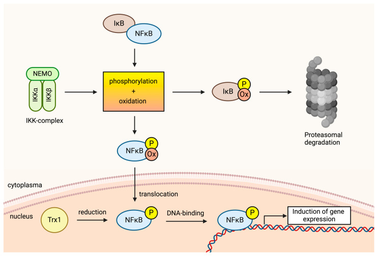
Reactive oxygen species (ROS) are central to inter- and intracellular signaling. Their localized and transient effects are due to their short half-life, especially when generated in controlled amounts. Upon T cell receptor (TCR) activation, regulated ROS signaling is primarily initiated by complexes I and III of the electron transport chain (ETC). Subsequent ROS production triggers the activation of nicotinamide adenine dinucleotide phosphate oxidase 2 (NADPH oxidase 2), prolonging the oxidative signal. This signal then engages kinase signaling cascades such as the mitogen-activated protein kinase (MAPK) pathway and increases the activity of REDOX-sensitive transcription factors such as nuclear factor-kappa B (NF-κB) and activator protein-1 (AP-1). To limit ROS overproduction and prevent oxidative stress, nuclear factor erythroid 2-related factor 2 (Nrf2) and antioxidant proteins such as superoxide dismutases (SODs) finely regulate signal intensity and are capable of terminating the oxidative signal when needed. Thus, oxidative signals, such as T cell activation, are well-controlled and critical for cellular communication.

Keywords: T cell activation; T cell receptor (TCR); T lymphocytes; electron transport chain (ETC); glycolysis; metabolic shift; oxidative signal; reactive oxygen species (ROS).


Conflict of interest statement

The authors declare no conflict of interest.

Figures

Figure 1
Figure 1
Schematic representation of oxidative signaling in T cells. Stimulation of the T cell receptor (TCR) leads to phosphorylation and activation of tyrosine kinase ZAP70, which phosphorylates LAT. Consequently, LAT recruits PLCγ1, which generates inositol 3,4,5-triphosphate (IP3) and diacylglycerol (DAG). At this point, the activation-induced signal diverges. IP3 is responsible for releasing Ca2+ into the cytosol. DAG activates the RAS guanyl-releasing protein 1 (RAS-GRP) and PKCθ. RAS-GRP activates Rat sarcoma (RAS) and subsequent kinase signaling. PKCθ induces ROS release via the mitochondria. Both signals are essential for the induction of activation-induced gene expression in T cells. Mitochondrial ROS release is a prerequisite for the induction of NADPH oxidase 2. ROS generation via NADPH oxidase 2 leads to a sustained oxidative signal. The figure was created with BioRender.com (accessed on 31 May 2024).
Figure 2
Figure 2
Complex I (CI) releases O2•− into the mitochondrial matrix, where most of it is converted to H2O2. However, certain conditions can lead to increased H2O2 production through other reactions. This high H2O2 production induces MnSOD, which further accelerates the conversion of O2•− to H2O2. As a result, fewer alternative reactions leading to H2O2 formation occur. Overall, this leads to reduced H2O2 production and a downregulation of the oxidative signal. The figure was created with BioRender.com.
Figure 3
Figure 3
After TCR stimulation, PLCγ1 generates 3,4,5-triphosphate (IP3) and diacylglycerol (DAG). IP3 induces Ca2+ influx into the cytosol and activates NF-AT via calcineurin. Simultaneously, DAG activates PKCθ. PKCθ most likely phosphorylates ADPGK. ADPGK induces a redirection of glycolysis, allowing electrons to be directly transferred to ubiquinone via GPD2. From there, electrons can be retrogradely directed towards Complex I of the ETC and forward directed to Complex III. At these complexes, ROS is released, activating REDOX-sensitive transcription factors and inducing the expression of specific genes essential for T cell activation. The figure was created with BioRender.com.
Figure 4
Figure 4
Overview of T cell subsets. CD8+ T cells, also known as cytotoxic T lymphocytes (CTLs), are a vital component of the adaptive immune system. Their primary function is to identify and destroy infected or malignant cells. CD4+ T cells, also known as helper T cells, are crucial for orchestrating the immune response. They assist other immune cells by releasing cytokines that regulate the activity, growth, and differentiation of various immune cells. CD4+ can further be divided into Th1 cells (expressing T-box expressed in T cells [T-bet]), Th2 cells (expressing the GATA binding protein 3 [GATA3]), Th17 cells (expressing the RAR-related orphan receptor gamma t [RORγt]), and T regulatory cells (Treg; expressing elevated levels of (Forkhead-Box-Protein P3 [FOXP3]). Th1 cells promote cell-mediated immunity by activating macrophages and cytotoxic T cells. They are crucial for defending against intracellular pathogens like viruses and certain bacteria. Th2 cells support humoral immunity by stimulating B cells to produce antibodies. They are important for combating extracellular parasites and allergens. Th17 cells are involved in defending against extracellular bacteria and fungi. They also play a role in autoimmune diseases by promoting inflammation. Tregs maintain immune tolerance by suppressing immune responses, preventing autoimmune diseases, and controlling inflammation. The figure was created with BioRender.com.
Figure 5
Figure 5
ROS-induced oxidation of free thiol groups activates MAP kinase kinase kinases (MAPKKKs), which phosphorylate MAPKK and, subsequently, MAPK. This signaling pathway is further amplified by the oxidation of MAPK phosphatases, which are inhibited by oxidation of a thiol residue in their active site, resulting in full activation of the MAPK cascade. OX = oxidation, P = phosphorylation. The figure was created with BioRender.com.
Figure 6
Figure 6
The role of oxidative signals in the activation of NF-κB. The inhibitor of NF-κB (IκB) is phosphorylated by the IκB kinase (IKK) complex. Additionally, a pro-oxidative environment in the cytosol causes IκB to become oxidized. Both phosphorylation and oxidation result in the accelerated degradation of IκB, so NF-κB is released. The free NF-κB can now translocate to the nucleus. Oxidation further accelerates this translocation. The oxidation of NF-κB is enabled by a shift in the cytosolic REDOX homeostasis towards a pro-oxidative state, induced by the oxidative signal. In the nucleus, NF-κB must be reduced again to enable optimal DNA binding. This is primarily achieved by Thioredoxin 1 (TRX1). The figure was created with BioRender.com 4.3.3. Nuclear Factor of Activated t cells (NF-AT).
Figure 7
Figure 7
After TCR stimulation, two signaling pathways are induced via 3,4,5-triphosphate (IP3) and diacylglycerol (DAG). The IP3-dependent pathway leads to Ca2+ release into the cytosol, activating NF-AT. The DAG-dependent pathway activates PKCθ and induces an oxidative signal via Complex I and III of the mitochondrial electron transport chain (ETC). Additionally, the MAPK signaling pathway is induced via PKCθ. The oxidative signal originating from mitochondrial ETC amplifies the MAPK signaling pathway and activates the redox-sensitive transcription factors AP1, NF-κB, and Nrf2. AP1 and NF-κB, together with NF-AT, create the minimal requirement for T cell activation. Conversely, Nrf2, by inducing antioxidant proteins, contributes to the control and potential termination of the oxidative signal. The figure was created with BioRender.com Oxidative signals in T cells are essential for the initiation of a T cell immune response. Several publications and data have addressed the induction of oxidative signals by TCR stimulation and the mechanisms by which these signals are generated. However, the role of co-stimulation in oxidative signaling is not well understood. This represents a knowledge gap that urgently needs to be investigated in detail.

Similar articles

Cited by

References

    1. Bendall D.S., Howe C.J., Nisbet E.G., Nisbet R.E.R. Photosynthetic and atmospheric evolution. Introduction. Philos. Trans. R. Soc. Lond. B Biol. Sci. 2008;363:2625–2628. doi: 10.1098/rstb.2008.0058. - DOI - PMC - PubMed
    1. Gumsley A.P., Chamberlain K.R., Bleeker W., Söderlund U., de Kock M.O., Larsson E.R., Bekker A. Timing and tempo of the Great Oxidation Event. Proc. Natl. Acad. Sci. USA. 2017;114:1811–1816. doi: 10.1073/pnas.1608824114. - DOI - PMC - PubMed
    1. Sessions A.L., Doughty D.M., Welander P.V., Summons R.E., Newman D.K. The continuing puzzle of the great oxidation event. Curr. Biol. 2009;19:R567–R574. doi: 10.1016/j.cub.2009.05.054. - DOI - PubMed
    1. West J.B. The strange history of atmospheric oxygen. Physiol. Rep. 2022;10:e15214. doi: 10.14814/phy2.15214. - DOI - PMC - PubMed
    1. Case A.J. On the Origin of Superoxide Dismutase: An Evolutionary Perspective of Superoxide-Mediated Redox Signaling. Antioxidants. 2017;6:82. doi: 10.3390/antiox6040082. - DOI - PMC - PubMed

Substances