. 2024 Jun 1;25(11):6120.
doi: 10.3390/ijms25116120.

Inhibition of SARS-CoV-2 Nsp9 ssDNA-Binding Activity and Cytotoxic Effects on H838, H1975, and A549 Human Non-Small Cell Lung Cancer Cells: Exploring the Potential of Nepenthes miranda Leaf Extract for Pulmonary Disease Treatment

Affiliations

Inhibition of SARS-CoV-2 Nsp9 ssDNA-Binding Activity and Cytotoxic Effects on H838, H1975, and A549 Human Non-Small Cell Lung Cancer Cells: Exploring the Potential of Nepenthes miranda Leaf Extract for Pulmonary Disease Treatment

Hsin-Hui Su et al. Int J Mol Sci. .

Abstract

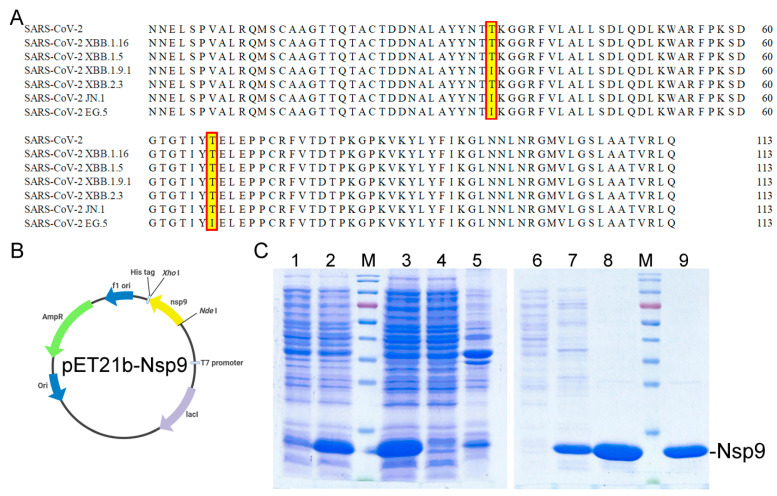
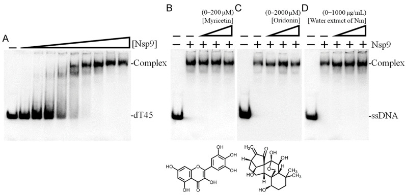
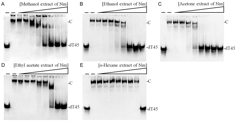
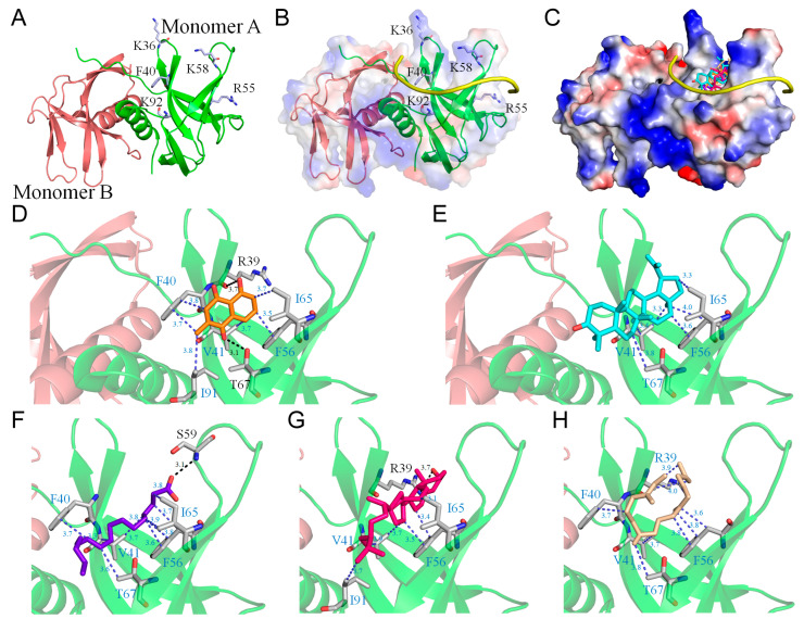
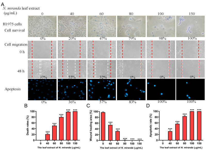
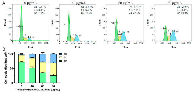
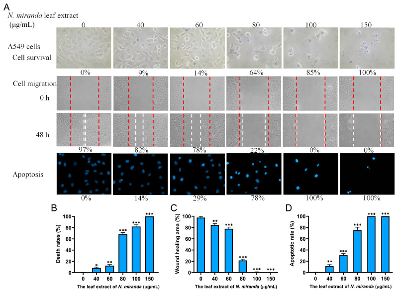
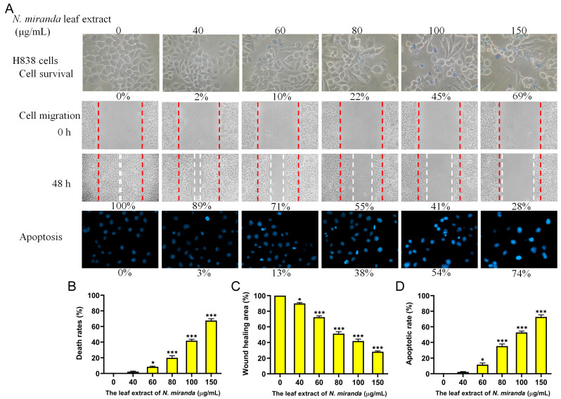
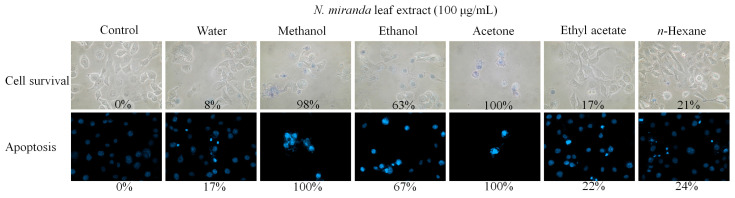
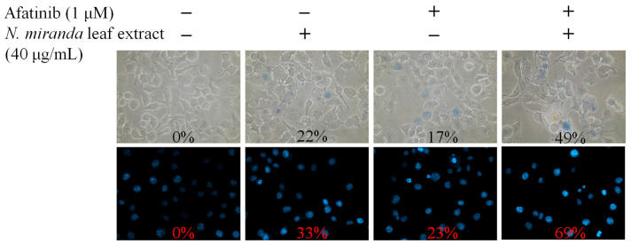
Carnivorous pitcher plants from the genus Nepenthes are renowned for their ethnobotanical uses. This research explores the therapeutic potential of Nepenthes miranda leaf extract against nonstructural protein 9 (Nsp9) of SARS-CoV-2 and in treating human non-small cell lung carcinoma (NSCLC) cell lines. Nsp9, essential for SARS-CoV-2 RNA replication, was expressed and purified, and its interaction with ssDNA was assessed. Initial tests with myricetin and oridonin, known for targeting ssDNA-binding proteins and Nsp9, respectively, did not inhibit the ssDNA-binding activity of Nsp9. Subsequent screenings of various N. miranda extracts identified those using acetone, methanol, and ethanol as particularly effective in disrupting Nsp9's ssDNA-binding activity, as evidenced by electrophoretic mobility shift assays. Molecular docking studies highlighted stigmast-5-en-3-ol and lupenone, major components in the leaf extract of N. miranda, as potential inhibitors. The cytotoxic properties of N. miranda leaf extract were examined across NSCLC lines H1975, A549, and H838, focusing on cell survival, apoptosis, and migration. Results showed a dose-dependent cytotoxic effect in the following order: H1975 > A549 > H838 cells, indicating specificity. Enhanced anticancer effects were observed when the extract was combined with afatinib, suggesting synergistic interactions. Flow cytometry indicated that N. miranda leaf extract could induce G2 cell cycle arrest in H1975 cells, potentially inhibiting cancer cell proliferation. Gas chromatography-mass spectrometry (GC-MS) enabled the tentative identification of the 19 most abundant compounds in the leaf extract of N. miranda. These outcomes underscore the dual utility of N. miranda leaf extract in potentially managing SARS-CoV-2 infection through Nsp9 inhibition and offering anticancer benefits against lung carcinoma. These results significantly broaden the potential medical applications of N. miranda leaf extract, suggesting its use not only in traditional remedies but also as a prospective treatment for pulmonary diseases. Overall, our findings position the leaf extract of N. miranda as a promising source of natural compounds for anticancer therapeutics and antiviral therapies, warranting further investigation into its molecular mechanisms and potential clinical applications.

Keywords: AntoDock; NSCLC; Nepenthes; Nsp9; SARS-CoV-2; anticancer; lupenone; plumbagin; stigmast-5-en-3-ol.


Conflict of interest statement

The authors declare no conflicts of interest.

Figures

Figure 1
Figure 1
Recombinant Nsp9 Protein from SARS-CoV-2. (A) Amino acid sequence alignment of Nsp9 from SARS-CoV-2. Sequence alignments across variants demonstrate high conservation with minor mutations (T35I and T67I) observed. (B) Construction of the expression plasmid pET21b-Nsp9. The synthesized gene, including NdeI and XhoI restriction sites, was ligated into the pET21b expression vector. (C) Protein expression and purification. Analysis was performed using 12% SDS-PAGE. The analysis includes bacterial culture before (lane 1) and after IPTG induction (lane 2), sonicated and centrifuged E. coli cells yielding supernatant (lane 3), supernatant applied to Ni-NTA column with analysis of flowthrough (lane 4), and pellet content (lane 5). Chromatographic steps with serial elutions in 5 mM imidazole buffer (20 mM Tris–HCl, 5 mM imidazole, and 0.5 M NaCl, pH 7.9; lane 6), 60 mM (lane 7), 100 mM (lane 8), and 200 mM (lane 9) imidazole were performed to obtain purified Nsp9. Elutions with 100 mM and 200 mM imidazole were effective in achieving homogeneity.
Figure 2
Figure 2
ssDNA binding activity of Nsp9. (A) Purified recombinant Nsp9 at various concentrations (0, 3, 6, 13, 25, 50, 75, 100, 150, 200 μM) was incubated with biotin-labeled ssDNA dT45 at 37 °C for 60 min. An increase in Nsp9 concentration resulted in a notable band shift, indicative of ssDNA binding. Binding constants ([Protein]50) were determined through linear interpolation based on the protein concentration. Binding assays were also conducted with the inclusion of (B) myricetin, (C) oridonin, or (D) water extract from N. miranda leaves in the protein solution, using 100 μM Nsp9. Myricetin and oridonin were dissolved in 10% dimethyl sulfoxide (DMSO). Nm denotes N. miranda.
Figure 3
Figure 3
Inhibition assay of Nsp9 ssDNA binding activity using N. miranda extracts. Nsp9 (100 μM) was incubated with N. miranda leaf extracts prepared with (A) methanol, (B) ethanol, (C) acetone, (D) ethyl acetate, and (E) n-hexane at concentrations ranging from 0 to 1000 μg/mL (0, 7.8, 15.6, 31.3, 62.5, 125, 250, 500, and 1000 μg/mL). Increasing concentrations of extracts led to an observable reduction in ssDNA band shift, indicating disruption of the Nsp9–ssDNA complex formation. Notably, the extracts obtained with methanol, ethanol, and acetone showed significant inhibitory effects on Nsp9 activity.
Figure 4
Figure 4
Molecular docking analysis of Nsp9. (A) The crystal structure of the Nsp9 dimer (PDB ID 6WXD), with monomers A and B colored differently. Key nucleic acid-binding residues K36, F40, R55, K58, and K92 are labeled for clarity. (B) A hypothetical model of the Nsp9–RNA complex, constructed based on the assumption that residues K36, F40, R55, K58, and K92 are crucial for nucleic acid binding. The RNA modeled in the complex is highlighted in yellow. (C) Docking analysis illustrating the five most abundant compounds from the N. miranda leaf extract individually docked into Nsp9: plumbagin (orange), lupenone (cyan), palmitic acid (purple-blue), stigmast-5-en-3-ol (hot pink), and neophytadiene (wheat). These compounds interact with RNA binding sites, occupying the cavity of the RNA-binding surface. Charge distribution patterns are shown to delineate RNA binding sites for enhanced clarity. (DH) depict the binding modes of plumbagin, lupenone, palmitic acid, stigmast-5-en-3-ol, and neophytadiene, respectively, to Nsp9. Each compound binds to Nsp9 with unique poses and at different sites. Residues involved in hydrogen bonding are marked in black, while those contributing to hydrophobic interactions are shown in blue.
Figure 5
Figure 5
Anticancer potential of N. miranda leaf extract on H1975 lung carcinoma cells. (A) The effect of N. miranda extract on cell survival, migration, and nuclear condensation in H1975 cells. (B) Trypan blue exclusion assay results demonstrating cell viability post-exposure to varying concentrations of N. miranda extract. (C) Wound-healing assay results showing the migration of H1975 cells before and 48 h after treatment with the extract at different concentrations. (D) Hoechst staining results depicting the levels of apoptosis and DNA fragmentation across a range of N. miranda extract concentrations. Statistical significance relative to the control is indicated by *** for p < 0.001.
Figure 6
Figure 6
(A) Alteration of cell cycle progression by N. miranda leaf extract in H1975 cells. H1975 cells underwent treatment with a control solution (0.1% DMSO) or with N. miranda leaf extract at specified concentrations for 24 h and were subsequently fixed in 70% ethanol overnight. The cells were then stained with propidium iodide (PI) for 30 min before analysis via flow cytometry. (B) The cell cycle distribution.
Figure 7
Figure 7
Anticancer potential of N. miranda leaf extract on A549 lung carcinoma cells. (A) The effect of N. miranda extract on cell survival, migration, and nuclear condensation in A549 cells. (B) Trypan blue exclusion assay results demonstrating cell viability post-exposure to varying concentrations of N. miranda extract. (C) Wound-healing assay results showing the migration of A549 cells before and 48 h after treatment with the extract at different concentrations. (D) Hoechst staining results depicting the levels of apoptosis and DNA fragmentation across a range of N. miranda extract concentrations. Statistical significance relative to the control is indicated by * for p < 0.05, ** for p < 0.01, and *** for p < 0.001.
Figure 8
Figure 8
Anticancer potential of N. miranda leaf extract on H838 lung carcinoma cells. (A) The effect of N. miranda extract on cell survival, migration, and nuclear condensation in H838 cells. (B) Trypan blue exclusion assay results demonstrating cell viability post-exposure to varying concentrations of N. miranda extract. (C) Wound-healing assay results showing the migration of H838 cells before and 48 h after treatment with the extract at different concentrations. (D) Hoechst staining results depicting the levels of apoptosis and DNA fragmentation across a range of N. miranda extract concentrations. Statistical significance relative to the control is indicated by * for p < 0.05 and *** for p < 0.001.
Figure 9
Figure 9
Anticancer potential of N. miranda leaf extract against H1975 lung carcinoma cells, based on solvent effects. The effect of extracts prepared using different solvents—distilled water, methanol, ethanol, acetone, ethyl acetate, and n-hexane—on cell survival and nuclear condensation, as assessed through trypan blue exclusion assay and Hoechst staining was examined. Extracts obtained with acetone and methanol demonstrated the highest anticancer potential among the tested solvents.
Figure 10
Figure 10
Synergistic effects of N. miranda leaf extract and afatinib on H1975 cells. The combined effect of N. miranda leaf extract (40 μg/mL) and afatinib (1 μM) on cell survival and apoptosis in H1975 cells was illustrated. Cell viability was assessed using trypan blue exclusion staining, and apoptosis was detected through Hoechst staining. The results suggest that combining afatinib with N. miranda leaf extract may enhance therapeutic efficacy against lung cancer.

Similar articles

References

    1. Wójciak M., Feldo M., Stolarczyk P., Płachno B.J. Carnivorous Plants from Nepenthaceae and Droseraceae as a Source of Secondary Metabolites. Molecules. 2023;28:2155. doi: 10.3390/molecules28052155. - DOI - PMC - PubMed
    1. Miclea I. Secondary Metabolites with Biomedical Applications from Plants of the Sarraceniaceae Family. Int. J. Mol. Sci. 2022;23:9877. doi: 10.3390/ijms23179877. - DOI - PMC - PubMed
    1. Tripathi S.K., Panda M., Biswal B.K. Emerging role of plumbagin: Cytotoxic potential and pharmaceutical relevance towards cancer therapy. Food Chem. Toxicol. 2019;125:566–582. doi: 10.1016/j.fct.2019.01.018. - DOI - PubMed
    1. Miguel S., Hehn A., Bourgaud F. Nepenthes: State of the art of an inspiring plant for biotechnologists. J. Biotechnol. 2018;265:109–115. doi: 10.1016/j.jbiotec.2017.11.014. - DOI - PubMed
    1. Guan H.-H., Huang Y.-H., Lin E.-S., Chen C.-J., Huang C.-Y. Plumbagin, a Natural Product with Potent Anticancer Activities, Binds to and Inhibits Dihydroorotase, a Key Enzyme in Pyrimidine Biosynthesis. Int. J. Mol. Sci. 2021;22:6861. doi: 10.3390/ijms22136861. - DOI - PMC - PubMed

MeSH terms