Lead Decreases Bone Morphogenetic Protein-7 (BMP-7) Expression and Increases Renal Cell Carcinoma Growth in a Sex-Divergent Manner
- PMID: 38892327
- PMCID: PMC11172964
- DOI: 10.3390/ijms25116139
Lead Decreases Bone Morphogenetic Protein-7 (BMP-7) Expression and Increases Renal Cell Carcinoma Growth in a Sex-Divergent Manner
Abstract
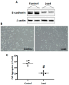
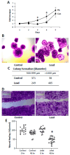
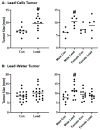
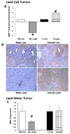
Both tissue and blood lead levels are elevated in renal cell carcinoma (RCC) patients. These studies assessed the impact of the subchronic lead challenge on the progression of RCC in vitro and in vivo. Lead challenge of Renca cells with 0.5 μM lead acetate for 10 consecutive passages decreased E-cadherin expression and cell aggregation. Proliferation, colony formation, and wound healing were increased. When lead-challenged cells were injected into mice, tumor size at day 21 was increased; interestingly, this increase was seen in male but not female mice. When mice were challenged with 32 ppm lead in drinking water for 20 weeks prior to tumor cell injection, there was an increase in tumor size in male, but not female, mice at day 21. To investigate the mechanism underlying the sex differences, the expression of sex hormone receptors in Renca cells was examined. Control Renca cells expressed estrogen receptor (ER) alpha but not ER beta or androgen receptor (AR), as assessed by qPCR, and the expression of ERα was increased in tumors in both sexes. In tumor samples harvested from lead-challenged cells, both ERα and AR were detected by qPCR, yet there was a significant decrease in AR seen in lead-challenged tumor cells from male mice only. This was paralleled by a plate-based array demonstrating the same sex difference in BMP-7 gene expression, which was also significantly decreased in tumors harvested from male but not female mice; this finding was validated by immunohistochemistry. A similar expression pattern was seen in tumors harvested from the mice challenged with lead in the drinking water. These data suggest that lead promotes RCC progression in a sex-dependent via a mechanism that may involve sex-divergent changes in BMP-7 expression.
Keywords: BMP-7; androgen receptor; estrogen receptor α; lead; renal cell carcinoma.
Conflict of interest statement
The authors declare no conflict of interest.
Figures


Similar articles
-
Estrogen inhibits renal cell carcinoma cell progression through estrogen receptor-β activation.PLoS One. 2013;8(2):e56667. doi: 10.1371/journal.pone.0056667. Epub 2013 Feb 27. PLoS One. 2013. PMID: 23460808 Free PMC article.
-
Androgen receptor decreases renal cell carcinoma bone metastases via suppressing the osteolytic formation through altering a novel circEXOC7 regulatory axis.Clin Transl Med. 2021 Mar;11(3):e353. doi: 10.1002/ctm2.353. Clin Transl Med. 2021. PMID: 33783995 Free PMC article.
-
Androgen receptor (AR) suppresses miRNA-145 to promote renal cell carcinoma (RCC) progression independent of VHL status.Oncotarget. 2015 Oct 13;6(31):31203-15. doi: 10.18632/oncotarget.4522. Oncotarget. 2015. PMID: 26304926 Free PMC article.
-
C1QBP Regulates YBX1 to Suppress the Androgen Receptor (AR)-Enhanced RCC Cell Invasion.Neoplasia. 2017 Feb;19(2):135-144. doi: 10.1016/j.neo.2016.12.003. Epub 2017 Jan 17. Neoplasia. 2017. PMID: 28107702 Free PMC article.
-
Intracrine androgen biosynthesis in renal cell carcinoma.Br J Cancer. 2017 Mar 28;116(7):937-943. doi: 10.1038/bjc.2017.42. Epub 2017 Mar 2. Br J Cancer. 2017. PMID: 28253524 Free PMC article.
References
-
- Surveillance Epidemiology and End Results. SEER Stat Fact Sheets. National Cancer Institute. [(accessed on 27 May 2024)]; Available online: https://seer.cancer.gov/statfacts/html/kidrp.html.
-
- McLaughlin J.K., Lipworth L., Tarone R.E., Blot W.J. Cancer Epidemiology and Prevention. Oxford University Press; Oxford, UK: 2006. pp. 1087–1100. Kidney cancer.
-
- Reilly R., Spalding S., Walsh B., Wainer J., Pickens S., Royster M., Villanacci J., Little B.B. Chronic environmental and occupational lead exposure and kidney function among African Americans: Dallas Lead Project II. Int. J. Environ. Res. Public Health. 2018;15:2875. doi: 10.3390/ijerph15122875. - DOI - PMC - PubMed
MeSH terms
Substances
Grants and funding
LinkOut - more resources
Full Text Sources
Medical
Research Materials

