. 2024 Jun 3;25(11):6149.
doi: 10.3390/ijms25116149.

Endogenous Hormone Levels and Transcriptomic Analysis Reveal the Mechanisms of Bulbil Initiation in Pinellia ternata

Affiliations

Endogenous Hormone Levels and Transcriptomic Analysis Reveal the Mechanisms of Bulbil Initiation in Pinellia ternata

Lan Mou et al. Int J Mol Sci. .

Abstract

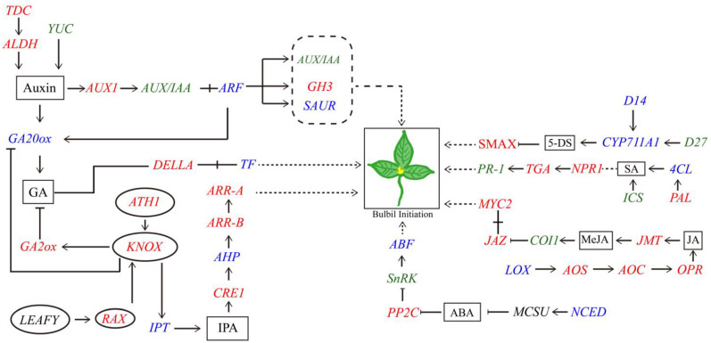
Pinellia ternata is a medicinal plant that has important pharmacological value, and the bulbils serve as the primary reproductive organ; however, the mechanisms underlying bulbil initiation remain unclear. Here, we characterized bulbil development via histological, transcriptomic, and targeted metabolomic analyses to unearth the intricate relationship between hormones, genes, and bulbil development. The results show that the bulbils initiate growth from the leaf axillary meristem (AM). In this stage, jasmonic acid (JA), abscisic acid (ABA), isopentenyl adenosine (IPA), and salicylic acid (SA) were highly enriched, while indole-3-acetic acid (IAA), zeatin, methyl jasmonate (MeJA), and 5-dexoxystrigol (5-DS) were notably decreased. Through OPLS-DA analysis, SA has emerged as the most crucial factor in initiating and positively regulating bulbil formation. Furthermore, a strong association between IPA and SA was observed during bulbil initiation. The transcriptional changes in IPT (Isopentenyltransferase), CRE1 (Cytokinin Response 1), A-ARR (Type-A Arabidopsis Response Regulator), B-ARR (Type-B Arabidopsis Response Regulator), AUX1 (Auxin Resistant 1), ARF (Auxin Response Factor), AUX/IAA (Auxin/Indole-3-acetic acid), GH3 (Gretchen Hagen 3), SAUR (Small Auxin Up RNA), GA2ox (Gibberellin 2-oxidase), GA20ox (Gibberellin 20-oxidase), AOS (Allene oxide synthase), AOC (Allene oxide cyclase), OPR (Oxophytodienoate Reductase), JMT (JA carboxy l Methyltransferase), COI1 (Coronatine Insensitive 1), JAZ (Jasmonate ZIM-domain), MYC2 (Myelocytomatosis 2), D27 (DWARF27), SMAX (Suppressor of MAX2), PAL (Phenylalanine Ammonia-Lyase), ICS (Isochorismate Synthase), NPR1 (Non-expressor of Pathogenesis-related Genes1), TGA (TGACG Sequence-specific Binding), PR-1 (Pathogenesis-related), MCSU (Molybdenium Cofactor Sulfurase), PP2C (Protein Phosphatase 2C), and SnRK (Sucrose Non-fermenting-related Protein Kinase 2) were highly correlated with hormone concentrations, indicating that bulbil initiation is coordinately controlled by multiple phytohormones. Notably, eight TFs (transcription factors) that regulate AM initiation have been identified as pivotal regulators of bulbil formation. Among these, WUS (WUSCHEL), CLV (CLAVATA), ATH1 (Arabidopsis Thaliana Homeobox Gene 1), and RAX (Regulator of Axillary meristems) have been observed to exhibit elevated expression levels. Conversely, LEAFY demonstrated contrasting expression patterns. The intricate expression profiles of these TFs are closely associated with the upregulated expression of KNOX(KNOTTED-like homeobox), suggesting a intricate regulatory network underlying the complex process of bulbil initiation. This study offers a profound understanding of the bulbil initiation process and could potentially aid in refining molecular breeding techniques specific to P. ternata.

Keywords: Pinellia ternata; bulbil; hormone; metabolomics; transcriptomics.


Conflict of interest statement

The funders had no role in the design of the study; in the collection, analyses, or interpretation of data; in the writing of the manuscript; or in the decision to publish the results.

Figures

Figure 1
Figure 1
Morphological characteristics of the top bulbil in different periods of development. Each column shows the same period of development for the top bulbil. BF, bulbil formation; BE, bulbil expansion; BM, bulbil maturity; EMC, external morphological characteristic of bulbil; 2D-V, 2D images of the bulbil transverse section; 2D-S, 2D image of bulbil longitudinal section; 3D, three-dimensional rendering image. EMC of BF, BE, and BM (ac); 2D-V of BF, BE, and BM (df); 2D-S of BF, BE, and BM (gi); 3D of BF, BE, and BM (jl).
Figure 2
Figure 2
Paraffin section pictures of SB and DB petioles at different growth stages of P. ternata. SB, single-bulbil type; DB, double-bulbil type; VB, vascular bundle; PC, parenchyma cell; PE, petiole epidermal cell. I, II, III, and IV represent the different development stages of SB and DB from germination to leaf spreading. The (a,d,g,h) morphological pictures of the top of the petiole and leaves of SB petioles in I, II, III, and IV stages, respectively; the (m,p,s,v) morphological pictures of the top of the petiole and leaves of DB petioles in I, II, III, and IV stages, respectively; (b,e,h,k,n,q,t,w) correspond to the paraffin section diagrams of the yellow-boxed areas within (a,d,g,j,m,p,s,v), respectively; (c,f,i,l,o,r,u,x) correspond to enlarged images of the yellow-boxed areas within (b,e,h,k,n,q,t,w), respectively. The red arrow shows where the top bulbil germinated in DB, while the green arrow indicates where no parenchymal hyperplasia occurred in SB during the same time.
Figure 3
Figure 3
Differentially accumulated metabolites in DUs and SUs. (A) PCA of the metabolites identified in DUs and SUs. (B) OPLS-DA plots and loading plots of DU vs. SU. (C) Permutation test of the OPLS-DA model for comparing SU and DU. (D) Volcano plot for DU vs. SU. SA, salicylic acid; IPA, isopentenyl adenosine; ABA, abscisic acid; JA, jasmonic acid; IAA, indole-3-acetic acid, 5-DS, 5-dexoxystrigol; MeJA, methyl jasmonate; SU, the top of the petiole in SB; DU, the top of the petiole in DB.
Figure 4
Figure 4
Analysis of differentially accumulated hormones in DU and SU groups. (A) HCA heatmap of DU vs. SU. The color blocks at different positions represent the relative expression of metabolites; red indicates high expression of the metabolite, and blue indicates low expression. (B) Matchstick analysis of DU vs. SU. The abscissa shows the log-transformed fold change, and the dot color represents the VIP value. **, 0.001 < p < 0.01; ***, p < 0.001. (C) The correlation analysis of DU vs. SU. The color blocks at different positions represent the magnitude of correlation coefficient between the metabolites, the red color indicates a positive correlation, and the blue color indicates a negative correlation. *, p < 0.05. SA, salicylic acid; IPA, isopentenyl adenosine; ABA, abscisic acid; JA, jasmonic acid; IAA, indole-3-acetic acid, 5-DS, 5-dexoxystrigol; MeJA, methyl jasmonate; SU, the top of the petiole in SB; DU, the top of the petiole in DB.
Figure 5
Figure 5
Transcriptome analysis of P. ternata. (A) Schematic representation of the transcriptomic sample positions: the top of the DB petiole, the middle of the DB petiole, the top of the SB petiole, and the middle of the SB petiole. (B) Unigene length distribution. (C) MDS Plot of DUs, SUs, DMs, SMs samples. (D) Classification of KEGG metabolic pathways. SU, the top of the petiole in SB; DU, the top of the petiole in DB; DM, the middle of the petiole in DB; SM, the middle of the petiole in SB; SB, single-bulbil type; DB, double-bulbil type.
Figure 6
Figure 6
DEGs. (A) Number of up- and downregulated genes. (B) Venn diagram of differentially expressed genes in different comparison groups. (C) A volcano plot was generated to visualize the DEGs identified from DU vs. SU comparison. (D) KEGG pathways enriched in DEGs were identified from DU vs. SU comparison. SU, the top of the petiole in SB; DU, the top of the petiole in DB; DM, the middle of petiole in DB; SM, the middle of petiole in SB.
Figure 7
Figure 7
Analysis of 5-DS, CTK, JA, ABA, IAA, GA, and SA biosynthesis pathways in DUs and SUs. The pinkish-purple and green colors in the upper heatmap reflect the upregulated and downregulated gene expressions, respectively. Similarly, the red and green colors in the lower heatmap represent increased and decreased metabolite accumulation, respectively. SA, salicylic acid; IPA, isopentenyl adenosine; ABA, abscisic acid; JA, jasmonic acid; IAA, indole-3-acetic acid, 5-DS, 5-dexoxystrigol; MeJA, methyl jasmonate; SU, the top of the petiole in SB; DU, the top of the petiole in DB.
Figure 8
Figure 8
Analysis of hormone signaling pathway components in DU and SU. The heatmap illustrates the expression levels of genes and metabolites involved in the IAA, CTK, ABA, JA, SA, and GA signaling pathways. The red and blue colors represent upregulated and downregulated gene expressions, respectively, while the yellow and blue colors reflect increased and decreased metabolite accumulations, respectively. The three columns on the left represent DU-1, DU-2, and DU-3, and the three columns on the right represent SU-1, SU-2, and SU-3. SU, the top of the petiole in SB; DU, the top of the petiole in DB.
Figure 9
Figure 9
Proposed regulatory network of bulbil initiation in P. ternata. Hormones are designated with black boxes, while genes regulating AM are denoted by oval shapes. Genes involved in hormone synthesis and signal transduction pathways exhibit upregulation in red, downregulation in green, and both upregulation and downregulation in blue. The dashed arrow with a pointed tip signifies the presumptive positive control relationship, the pointed arrow represents positive control, and the flat arrow line with a blunt tip indicates an inhibitory effect.
Figure 10
Figure 10
Overview of SB and DB in P. ternata. The rows indicate the top of the petiole. SB, single-bulbil type; DB, double-bulbil type. White arrowheads indicate different phenotypes at the top of the petiole with SB and DB. There is a bulbil at the top of the DB petiole, but none at the top of the SB petiole.

Similar articles

Cited by

References

    1. Duan X., Chen L., Liu Y., Chen H., Wang F., Hu Y. Integrated physicochemical, hormonal, and transcriptomic analysis reveals the underlying mechanism of callus formation in Pinellia ternata hydroponic cuttings. Front. Plant Sci. 2023;14:1189499. doi: 10.3389/fpls.2023.1189499. - DOI - PMC - PubMed
    1. Bai J., Qi J., Yang L., Wang Z., Wang R. A comprehensive review on ethnopharmacological, phytochemical, pharmacological and toxicological evaluation, and quality control of Pinellia ternata (Thunb.) Breit. J. Ethnopharmacol. 2022;298:115650. doi: 10.1016/j.jep.2022.115650. - DOI - PubMed
    1. Guo C., Li J., Li M., Xu X., Chen Y., Chu J., Yao X. Regulation Mechanism of Exogenous Brassinolide on Bulbil Formation and Development in Pinellia ternata. Front. Plant Sci. 2022;12:809769. doi: 10.3389/fpls.2021.809769. - DOI - PMC - PubMed
    1. Mao R., He Z. Pinellia ternata (Thunb.) Breit: A review of its germplasm resources, genetic diversity and active components. J. Ethnopharmacol. 2020;263:113252. doi: 10.1016/j.jep.2020.113252. - DOI - PubMed
    1. Xu W., Fan H., Pei X., Hua X., Xu T., He Q. mRNA-Seq and miRNA-Seq Analyses Provide Insights into the Mechanism of Pinellia ternata Bulbil Initiation Induced by Phytohormones. Genes. 2023;14:1727. doi: 10.3390/genes14091727. - DOI - PMC - PubMed