. 2024 Jun 4;25(11):6177.
doi: 10.3390/ijms25116177.

Contribution of Extracellular Particles Isolated from Morus sp. (Mulberry) Fruit to Their Reported Protective Health Benefits: An In Vitro Study

Affiliations

Contribution of Extracellular Particles Isolated from Morus sp. (Mulberry) Fruit to Their Reported Protective Health Benefits: An In Vitro Study

Neve R Garrett et al. Int J Mol Sci. .

Abstract

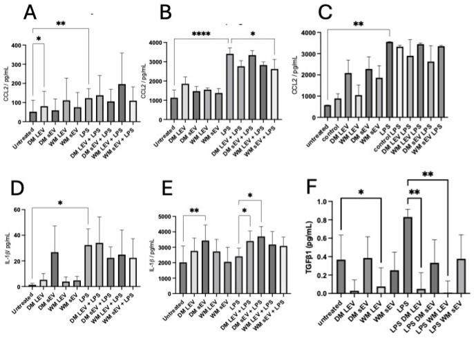
Morus sp. (mulberry) has a long tradition of use as a medicinal treatment, including for cardiovascular disease and type 2 diabetes, being shown to have antioxidant properties and to promote wound healing. Extracellular vesicles (EVs) are sub-micron, membrane-enclosed particles that were first identified in mammalian bodily fluids. EV-like particles have been described in plants (PDVs) and shown to have similar characteristics to mammalian EVs. We hypothesised that some of the health benefits previously attributed to the fruit of Morus sp. could be due to the release of PDVs. We isolated PDVs from Morus nigra and Morus alba via ultracentrifugation and incubated THP-1 monocytes, differentiated THP-1 macrophages, or HMEC-1 endothelial cells with pro-oxidant compounds DMNQ (THP-1) and glucose oxidase (HMEC-1) or lipopolysaccharide (LPS) in the presence of different fractions of mulberry EVs. Mulberry EVs augmented ROS production with DMNQ in THP-1 and caused the downregulation of ROS in HMEC-1. Mulberry EVs increased LPS-induced IL-1β secretion but reduced CCL2 and TGF-β secretion in THP-1 macrophages. In scratch wound assays, mulberry EVs inhibited HMEC-1 migration but increased proliferation in both low and high serum conditions, suggesting that they have opposing effects in these two important aspects of wound healing. One of the limitations of plant-derived therapeutics has been overcoming the low bioavailability of isolated compounds. We propose that PDVs could provide the link between physiological dose and therapeutic benefit by protecting plant active compounds in the GIT as well as potentially delivering genetic material or proteins that contribute to previously observed health benefits.

Keywords: Morus alba; Morus nigra; extracellular vesicles; inflammation; mulberry; oxidative stress; proliferation.


Conflict of interest statement

The authors declare no conflicts of interest.

Figures

Figure 1
Figure 1
Identification of different PDV fractions from dark and white mulberry fruit. LEVs and sEVs were isolated sequentially from dried DM (A,C,D,E) and WM (B,F,G,H). Representative flow cytometry of LEVs (C,F), blue represents total LEVs, Pink represents Annexin V+ LEVs, red repreents enumeration beads, NTA of sEVs (D,G), and NTA of control fraction (E,H).
Figure 2
Figure 2
Mulberry EVs modulate ROS production in human cells in vitro. THP-1 monocytes (A,D) PMA-differentiated macrophages (B,E) and HMEC-1 (C,F) were pre-loaded with dihydrorhodamine-1,2,3 and treated with DMNQ (monocytes and macrophages) or GO (HMEC-1) in the presence or absence of DM EVs (AC) or WM EVs (DF). All treatments were in triplicate, n = 4 isolations of mulberry EVs. Repeated measures of one-way ANOVA followed by Fisher’s LSD. * p < 0.05, ** p < 0.01, and **** p < 0.0001.
Figure 3
Figure 3
Mulberry EV modulation of LPS-stimulated cytokine secretion from THP-1 and HMEC-1. THP-1 monocytes (A,D), PMA-stimulated macrophages (B,E), and HMEC-1 (C) were treated with LPS in the presence or absence of mulberry EVs. The supernatants were assayed for CCL2 (AC), IL-1β (D,E), and TGFβ1 (F). All treatments were measured in triplicate, n = 4 isolations of mulberry EV. Repeated measures of one-way ANOVA followed by Fisher’s LSD. * p < 0.05, ** p < 0.01, and **** p < 0.0001.
Figure 4
Figure 4
Migration of HMEC-1 endothelial cells is inhibited by mulberry EVs in an in vitro scratch (wound healing) assay. HMEC-1 were seeded onto 24-well plates at a sufficient density to form a confluent monolayer. After performing a “scratch”, whole wells were immediately imaged using a Tecan Spark Cyto imaging plate reader, [2× magnification], and again after 18 h, culture in either 1% or 10% EV-depleted FBS, in the presence or absence of mulberry EVs. (A) Representative images of scratches in 1% and 10% FBS at T0 and T = 18 h. (B) Averages of % change in confluency for wells incubated in 1% FBS. (C) Averages of % change in confluency for wells incubated in 10% FBS, n = 4. Repeated measures of one-way ANOVA followed by Fisher’s LSD. ** p < 0.01, and *** p < 0.001. n.s. not significant.
Figure 5
Figure 5
Proliferation of HMEC-1 is enhanced by mulberry EVs in low- (A) and high- (10%) FBS-containing medium (B). Cells were seeded at a low density in 96-well plates. The following morning, they were treated with 1% or 10% FBS in the presence or absence of mulberry EVs. Proliferation was measured after the addition of Alamar Blue. Red dotted lines depict level of proliferation in cells treated with 1% FBS only. One-way ANOVA followed by Fisher’s LSD; n = 4. * p < 0.05, ** p < 0.01, and **** p < 0.0001.

Similar articles

Cited by

References

    1. Gundogdu M., Muradoglu F., Sensoy R.I.G., Yilmaz H. Determination of fruit chemical properties of Morus nigra L., Morus alba L. and Morus rubra L. by HPLC. Sci. Hortic. 2011;132:37–41. doi: 10.1016/j.scienta.2011.09.035. - DOI
    1. Rodrigues E.L., Marcelino G., Silva G.T., Figueiredo P.S., Garcez W.S., Corsino J., Guimarães R.C.A., Freitas K.C. Nutraceutical and Medicinal Potential of the Morus species in Metabolic Dysfunctions. Int. J. Mol. Sci. 2019;20:301. doi: 10.3390/ijms20020301. - DOI - PMC - PubMed
    1. Sánchez-Salcedo E.M., Sendra E., Carbonell-Barrachina Á., Martínez J.J., Hernández F. Fatty acids composition of Spanish black (Morus nigra L.) and white (Morus alba L.) mulberries. Food Chem. 2016;190:566–571. doi: 10.1016/j.foodchem.2015.06.008. - DOI - PubMed
    1. Sezai Ercisli and Emine O. Chemical composition of white (Morus alba), red (Morus rubra) and black (Morus nigra) mulberry fruits. Food Chem. 2007;103:1380–1384. doi: 10.1016/j.foodchem.2006.10.054. - DOI
    1. Jan B., Parveen R., Zahiruddin S., Khan M.U., Mohapatra S., Ahmad S. Nutritional constituents of mulberry and their potential applications in food and pharmaceuticals: A review. Saudi J. Biol. Sci. 2021;28:3909–3921. doi: 10.1016/j.sjbs.2021.03.056. - DOI - PMC - PubMed

LinkOut - more resources