Review
. 2024 Jun 4;25(11):6180.
doi: 10.3390/ijms25116180.

Endothelial-to-Mesenchymal Transition in Cardiovascular Pathophysiology

Affiliations
Review

Endothelial-to-Mesenchymal Transition in Cardiovascular Pathophysiology

Aman Singh et al. Int J Mol Sci. .

Abstract

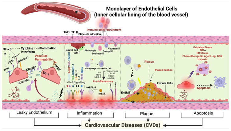
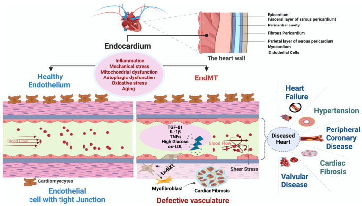
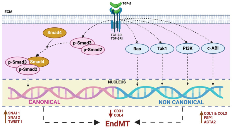
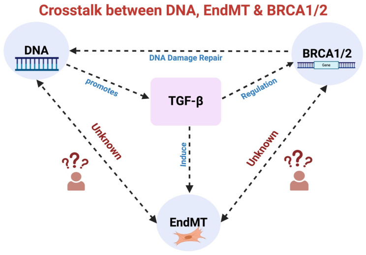
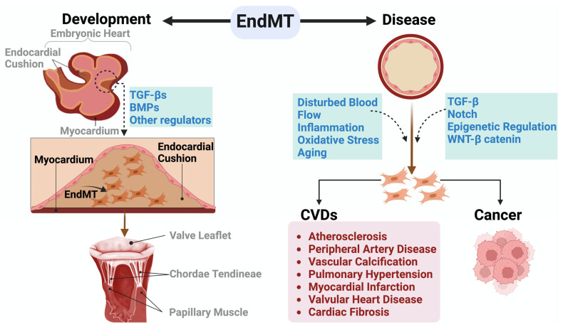
Under different pathophysiological conditions, endothelial cells lose endothelial phenotype and gain mesenchymal cell-like phenotype via a process known as endothelial-to-mesenchymal transition (EndMT). At the molecular level, endothelial cells lose the expression of endothelial cell-specific markers such as CD31/platelet-endothelial cell adhesion molecule, von Willebrand factor, and vascular-endothelial cadherin and gain the expression of mesenchymal cell markers such as α-smooth muscle actin, N-cadherin, vimentin, fibroblast specific protein-1, and collagens. EndMT is induced by numerous different pathways triggered and modulated by multiple different and often redundant mechanisms in a context-dependent manner depending on the pathophysiological status of the cell. EndMT plays an essential role in embryonic development, particularly in atrioventricular valve development; however, EndMT is also implicated in the pathogenesis of several genetically determined and acquired diseases, including malignant, cardiovascular, inflammatory, and fibrotic disorders. Among cardiovascular diseases, aberrant EndMT is reported in atherosclerosis, pulmonary hypertension, valvular disease, fibroelastosis, and cardiac fibrosis. Accordingly, understanding the mechanisms behind the cause and/or effect of EndMT to eventually target EndMT appears to be a promising strategy for treating aberrant EndMT-associated diseases. However, this approach is limited by a lack of precise functional and molecular pathways, causes and/or effects, and a lack of robust animal models and human data about EndMT in different diseases. Here, we review different mechanisms in EndMT and the role of EndMT in various cardiovascular diseases.

Keywords: EndMT; cardiovascular diseases; endothelial-to-mesenchymal transition; mechanisms.


Conflict of interest statement

The authors declare no conflicts of interest.

Figures

Figure 1
Figure 1
Endothelium and CVDs. Different mechanisms leading to leaky vasculature, increased inflammation, plaque development, or apoptosis in CVDs. The figure provides a detailed depiction of the interconnected mechanisms within endothelial dysfunction and their relevance to CVDs. It illustrates how the endothelium plays a crucial role in the pathogenesis of CVDs through various processes. The sequence begins with endothelial dysfunction leading to a leaky vasculature and increased vascular permeability, facilitated by TLR4 activation of the NF-κB pathway, upregulation of ICAM-1, VCAM-1, Selectins and subsequent cytokine release—all contributing to heightened inflammation. This cascade of events culminates in inflammation, affecting claudin-1 and ZO-1. The progression of inflammation exacerbates plaque development through the transformation of macrophages into foam cells, releasing pro-inflammatory cytokines, including IL-6, TNF-α, and MMP-7. Reduced phagocytosis and decreased CD163 expression highlight the impact of these processes on plaque instability within the vasculature. Additionally, upregulated vWF mediates endothelial activation and platelet adhesion, crucial for the inflammatory response. Moreover, vWF facilitates immune cell recruitment and acts as a signaling molecule, amplifying the inflammatory cascade. Plaque formation and rupture are highlighted as critical events triggered by endothelial dysfunction and oxLDL accumulation, contributing to the progression of CVDs. Finally, the figure illustrates apoptosis induced by various factors such as oxidative stress, hypoxia, reduced NO, ER stress, and chemotherapeutic agents and shear stress (as shown by red arrows pointing in the same direction, while others veer left or right, and some proceeding straight, capturing the dynamic nature of biomechanical forces acting on the endothelium). These factors contribute to apoptotic cell death within the vascular environment, emphasizing the diverse mechanisms through which endothelial dysfunction can lead to adverse outcomes in cardiovascular health, ultimately impacting the development and progression of various CVDs. TLR4: Toll-like Receptor 4, ICAM-1: Intercellular Adhesion Molecule-1, VCAM-1: Vascular Cell Adhesion Molecule-1, NF-κB: Nuclear Factor Kappa B, IL-6: Interleukin-6, TNF-α: Tumor Necrosis Factor- α, MMP-7: Matrix Metalloproteinase-7, CD163: Cluster of Differentiation 163, vWF: von Willebrand Factor, NO—Nitric Oxide, ER—Endoplasmic Reticulum, oxLDL: Oxidized Low-Density Lipoprotein, ZO-1: Zonula Occludens-1, EndMT: Endothelial-to-Mesenchymal Transition.
Figure 2
Figure 2
Figure depicting healthy endothelium and EndMT leading to different CVDs. Figure showing EndMT within endocardial endothelial cells, elucidating the contrast between the impermeable vascular structure of healthy endothelium and the disruptive impact of EndMT. Conversely, the EndMT, ignited by factors like shear stress, TGF-β, IL-1β, TNF-α, high glucose levels, and oxLDL, drives the transformation of active endothelial cells into myofibroblasts, promoting cardiac fibrosis. Beyond these factors, inflammation, aging, mitochondrial dysfunction, and autophagic dysfunction also contribute to the EndMT process. The appearance of a leaky endothelium signals the initiation of endothelial dysfunction within the endocardium, indicating a critical juncture in the path toward CVDs. This transformative journey not only disrupts endothelial integrity but heightens the risks of severe cardiovascular ailments such as heart failure, hypertension, peripheral coronary artery disease, cardiac fibrosis, and valvular abnormalities. These outcomes emphasize the intricate interplay between endothelial dysfunction, EndMT, and the spectrum of cardiovascular disorders, underscoring the necessity for a profound comprehension of these processes to drive targeted interventions and preserve cardiovascular well-being. EndMT: Endothelial-to-Mesenchymal Transition, TGF-β: Transforming Growth Factor-β, IL-1β: Interleukin-1β, TNF-α: Tumor Necrosis Factor-α, oxLDL: Oxidized Low-Density Lipoprotein.
Figure 3
Figure 3
Canonical and noncanonical TGF- β signaling in EndMT. TGF-β homodimers interact with the TGF-β I/II receptor complex on the cell surface to initiate the canonical TGF-β pathway. This triggers the phosphorylation and activation of Smad2 and Smad3 proteins. Subsequently, the activated Smad2/3 complex with Smad4 translocate to the nucleus and activate transcription of genes involved in creating profibrotic extracellular matrix components such as Col4, Col1, Col3 and transcription factors like Snai1, Snai2, and Twist1. Noncanonical pathways involving kinases PI3K, Tak1, Ras, and c-Abl also influence EndMT. These pathways can lead to different cellular responses, independent of Smad2/3 activation, either by decreasing endothelial-specific gene transcription or increasing mesenchymal-specific gene expression. These pathways impact the expression of genes associated with EndMT, leading to variations in the transcription of markers like CD31, Col4, Col1, Col3, FSP1, and Acta2. Notably, these effects are also influenced by transcription factors such as Snai1, Snai2, and Twist1. TGF-β: Transforming Growth Factor-β, ECM: Extracellular Matrix, TGF-βRI/II: Transforming Growth Factor-β Receptor I/II, EndMT: Endothelial-to-Mesenchymal Transition, Col4: Collagen Type IV, Col1: Collagen Type I, Col3: Collagen Type III, PI3K: Phosphatidylinositol 3-Kinase, Tak1: TGF-β-activated kinase 1, c-Abl: Abelson Tyrosine-Protein Kinase 1, CD31: Cluster of Differentiation 31, FSP1: Fibroblast-Specific Protein 1, Acta2: Alpha Smooth Muscle Actin, Snail1: Zinc Finger Protein SNAI1, Snail2: Zinc Finger Protein SNAI2, Twist: Twist Family BHLH Transcription Factor 1.
Figure 4
Figure 4
Unraveling the intricate interactions: TGF-β signaling, DNA damage repair, BRCA1/BRCA2, and EndMT. TGF-β signaling influences BRCA1/2 expression, pivotal in DNA damage repair, while DNA damage reciprocally enhances TGF-β signaling. Furthermore, TGF-β also induces EndMT. An established relationship exists between TGF-β and EndMT, TGF-β and DNA damage, and TGF-β and BRCA1/2. However, the direct effect of DNA damage and DNA damage repair molecules BRCA1/2 on EndMT remains unknown. TGF-β: Transforming Growth Factor-β, BRCA1/2: Breast Cancer Susceptibility Gene 1/2, EndMT: Endothelial-to-Mesenchymal Transition, DNA: Deoxyribonucleic Acid.
Figure 5
Figure 5
EndMT in development and disease. During embryonic development, specific endothelial cells within the AV canal engage in EndMT, facilitating the creation of endocardial cushions essential for the proper development of the AV valve. As the embryo matures, these endocardial cushions undergo remodeling to form durable valve leaflets and the supporting chordae tendineae. In adult life, EndMT plays a crucial role in the initiation and progression of various CVDs and cancer. EndMT: Endothelial-to-Mesenchymal Transition, CVDs: Cardiovascular Diseases.

References

    1. Carmeliet P., Jain R.K. Molecular mechanisms and clinical applications of angiogenesis. Nature. 2011;473:298–307. doi: 10.1038/nature10144. - DOI - PMC - PubMed
    1. Cybulsky M.I., Gimbrone M.A. Endothelial Expression of a Mononuclear Leukocyte Adhesion Molecule During Atherogenesis. Science. 1991;251:788–791. doi: 10.1126/science.1990440. - DOI - PubMed
    1. Glass C.K., Witztum J.L. Atherosclerosis. the Road Ahead. Cell. 2001;104:503–516. doi: 10.1016/S0092-8674(01)00238-0. - DOI - PubMed
    1. Rajendran P., Rengarajan T., Thangavel J., Nishigaki Y., Sakthisekaran D., Sethi G., Nishigaki I. The Vascular Endothelium and Human Diseases. Int. J. Biol. Sci. 2013;9:1057–1069. doi: 10.7150/ijbs.7502. - DOI - PMC - PubMed
    1. Li B., Syed M.H., Khan H., Singh K.K., Qadura M. The Role of Fatty Acid Binding Protein 3 in Cardiovascular Diseases. Biomedicines. 2022;10:2283. doi: 10.3390/biomedicines10092283. - DOI - PMC - PubMed

MeSH terms