

Semaglutide May Ameliorate Fibrosis and Inhibit Epithelial-Mesenchymal Transition in Intrauterine Adhesion Models
- PMID: 38892384
- PMCID: PMC11172622
- DOI: 10.3390/ijms25116196
Semaglutide May Ameliorate Fibrosis and Inhibit Epithelial-Mesenchymal Transition in Intrauterine Adhesion Models
Abstract
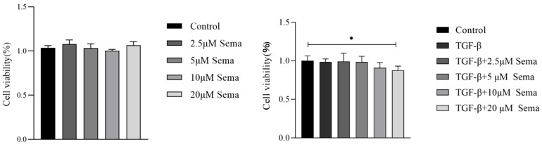
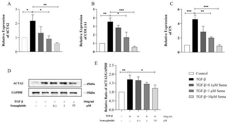
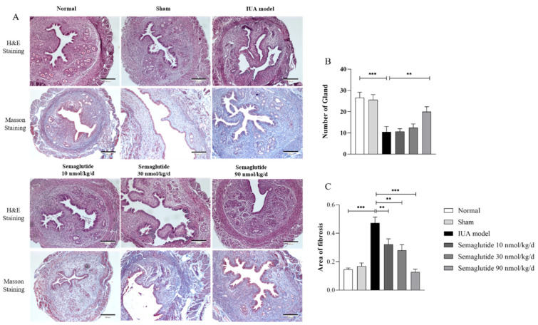
The purpose of this study was to explore the effect of Semaglutide on intrauterine adhesions and discover new drugs for such adhesions. In this study, the cell model was simulated by TGF-β1-induced human endometrial epithelial cells, and the animal model was established through mechanical curettage and inflammatory stimulation. After co-culturing with TGF-β1 with or without different concentrations of Semaglutide for 48 h, cells were collected for RT-qPCR and Western blotting analyses. Three doses were subcutaneously injected into experimental mice once a day for two weeks, while the control group received sterile ddH2O. The serum and uterine tissues of the mice were collected. HE and Masson staining were used for the uterine histomorphological and pathological analyses. RT-qPCR and Western blotting were used for mRNA and protein expression analyses. Serum indicators were detected using ELISA kits. The results showed that Semaglutide significantly reduced the mRNA levels of fibrosis indicators ACTA2, COL1A1, and FN and inflammatory indicators TNF-α, IL-6, and NF-κB in the two models. Semaglutide improved endometrium morphology, increased the number of endometrial glands, and reduced collagen deposition in IUA mice. The results also showed that Semaglutide could inhibit vimentin, E-Cadherin, and N-Cadherin in the two models. In summary, Semaglutide can ameliorate fibrosis and inflammation of intrauterine adhesions as well as inhibit epithelial-mesenchymal transition in IUA models.
Keywords: Semaglutide; epithelial–mesenchymal transition; fibrosis; intrauterine adhesions.
Conflict of interest statement
The authors declare no conflicts of interest.
Figures


Similar articles
-
Mechanism of electroacupuncture in treating uterine endometrial fibrosis in intrauterine adhesions rats based on Wnt/β-catenin pathway-mediated epithelial-mesenchymal transition.Zhen Ci Yan Jiu. 2024 Jun 25;49(6):566-576. doi: 10.13702/j.1000-0607.20240101. Zhen Ci Yan Jiu. 2024. PMID: 38897800 Chinese, English.
-
The regulatory effect of electroacupuncture on endometrial M1-type macrophages in rats with intrauterine adhesions.Zhen Ci Yan Jiu. 2024 May 25;49(5):487-498. doi: 10.13702/j.1000-0607.20230874. Zhen Ci Yan Jiu. 2024. PMID: 38764120 Chinese, English.
-
The glucagon-like peptide-1 (GLP-1) analog exenatide ameliorates intrauterine adhesions in mice.Peptides. 2021 Mar;137:170481. doi: 10.1016/j.peptides.2020.170481. Epub 2021 Jan 12. Peptides. 2021. PMID: 33450323
-
Recent Advances in Understandings Towards Pathogenesis and Treatment for Intrauterine Adhesion and Disruptive Insights from Single-Cell Analysis.Reprod Sci. 2021 Jul;28(7):1812-1826. doi: 10.1007/s43032-020-00343-y. Epub 2020 Oct 30. Reprod Sci. 2021. PMID: 33125685 Free PMC article. Review.
-
Transforming growth factor-β1 in intrauterine adhesion.Am J Reprod Immunol. 2020 Aug;84(2):e13262. doi: 10.1111/aji.13262. Epub 2020 Jun 3. Am J Reprod Immunol. 2020. PMID: 32379911 Review.
Cited by
-
The Effects of Oral Semaglutide on Hepatic Fibrosis in Subjects with Type 2 Diabetes in Real-World Clinical Practice: A Post Hoc Analysis of the Sapporo-Oral SEMA Study.Pharmaceuticals (Basel). 2025 Jan 19;18(1):129. doi: 10.3390/ph18010129. Pharmaceuticals (Basel). 2025. PMID: 39861190 Free PMC article.
-
Current Therapeutic Landscape for Metabolic Dysfunction-Associated Steatohepatitis.Int J Mol Sci. 2025 Feb 19;26(4):1778. doi: 10.3390/ijms26041778. Int J Mol Sci. 2025. PMID: 40004240 Free PMC article. Review.
-
Semaglutide enhances PINK1/Parkin‑dependent mitophagy in hypoxia/reoxygenation‑induced cardiomyocyte injury.Mol Med Rep. 2025 May;31(5):111. doi: 10.3892/mmr.2025.13476. Epub 2025 Feb 28. Mol Med Rep. 2025. PMID: 40017118 Free PMC article.
-
Live birth following multimodal therapy in a patient with asherman's syndrome, recurrent pregnancy loss, and polycystic ovarian syndrome: a case report and literature review.Contracept Reprod Med. 2025 Aug 7;10(1):45. doi: 10.1186/s40834-025-00384-1. Contracept Reprod Med. 2025. PMID: 40770747 Free PMC article.
References
-
- Hooker A.B., Lemmers M., Thurkow A.L., Heymans M.W., Opmeer B.C., Brölmann H.A., Mol B.W., Huirne J.A. Systematic review and meta-analysis of intrauterine adhesions after miscarriage: Prevalence, risk factors and long-term reproductive outcome. Hum. Reprod. Update. 2014;20:262–278. doi: 10.1093/humupd/dmt045. - DOI - PubMed
MeSH terms
Substances
Grants and funding
LinkOut - more resources
Full Text Sources
Research Materials
Miscellaneous
