

Molecular Force Sensors for Biological Application
- PMID: 38892386
- PMCID: PMC11173168
- DOI: 10.3390/ijms25116198
Molecular Force Sensors for Biological Application
Abstract
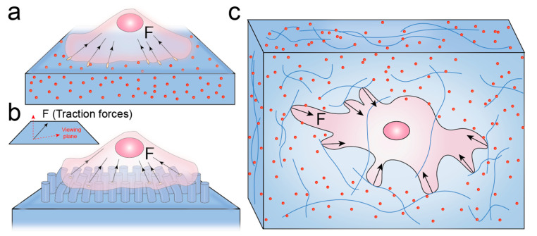
The mechanical forces exerted by cells on their surrounding microenvironment are known as cellular traction forces. These forces play crucial roles in various biological processes, such as tissue development, wound healing and cell functions. However, it is hard for traditional techniques to measure cellular traction forces accurately because their magnitude (from pN to nN) and the length scales over which they occur (from nm to μm) are extremely small. In order to fully understand mechanotransduction, highly sensitive tools for measuring cellular forces are needed. Current powerful techniques for measuring traction forces include traction force microscopy (TFM) and fluorescent molecular force sensors (FMFS). In this review, we elucidate the force imaging principles of TFM and FMFS. Then we highlight the application of FMFS in a variety of biological processes and offer our perspectives and insights into the potential applications of FMFS.
Keywords: cellular traction forces; fluorescent molecular force sensors; mechanotransduction; traction force microscopy.
Conflict of interest statement
The authors declare that they have no known competing financial interests or personal relationships that could have appeared to influence the work reported in this paper.
Figures


References
-
- Qi Y.-X., Yao Q.-P., Huang K., Shi Q., Zhang P., Wang G.-L., Han Y., Bao H., Wang L., Li H.-P., et al. Nuclear envelope proteins modulate proliferation of vascular smooth muscle cells during cyclic stretch application. Proc. Natl. Acad. Sci. USA. 2016;113:5293–5298. doi: 10.1073/pnas.1604569113. - DOI - PMC - PubMed
-
- Qi Y.-X., Jiang J., Jiang X.-H., Wang X.-D., Ji S.-Y., Han Y., Long D.-K., Shen B.-R., Yan Z.-Q., Chien S., et al. PDGF-BB and TGF-β1 on cross-talk between endothelial and smooth muscle cells in vascular remodeling induced by low shear stress. Proc. Natl. Acad. Sci. USA. 2011;108:1908–1913. doi: 10.1073/pnas.1019219108. - DOI - PMC - PubMed
Publication types
MeSH terms
LinkOut - more resources
Full Text Sources
Other Literature Sources
