Review
. 2024 Jun 4;25(11):6199.
doi: 10.3390/ijms25116199.

The Skin-Brain Axis: From UV and Pigmentation to Behaviour Modulation

Affiliations
Review

The Skin-Brain Axis: From UV and Pigmentation to Behaviour Modulation

Anna A Ascsillán et al. Int J Mol Sci. .

Abstract

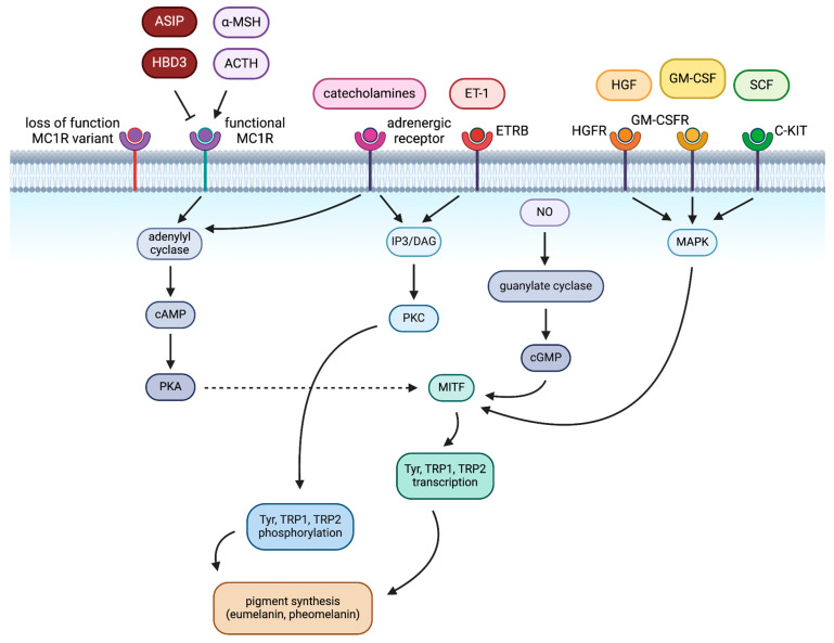
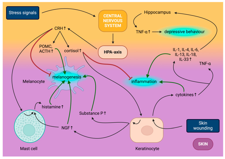
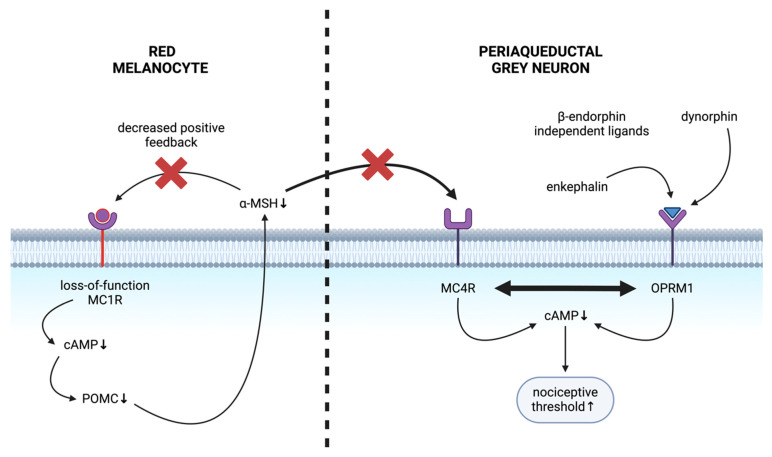
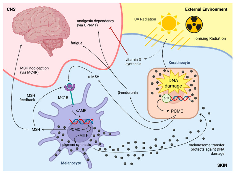
The skin-brain axis has been suggested to play a role in several pathophysiological conditions, including opioid addiction, Parkinson's disease and many others. Recent evidence suggests that pathways regulating skin pigmentation may directly and indirectly regulate behaviour. Conversely, CNS-driven neural and hormonal responses have been demonstrated to regulate pigmentation, e.g., under stress. Additionally, due to the shared neuroectodermal origins of the melanocytes and neurons in the CNS, certain CNS diseases may be linked to pigmentation-related changes due to common regulators, e.g., MC1R variations. Furthermore, the HPA analogue of the skin connects skin pigmentation to the endocrine system, thereby allowing the skin to index possible hormonal abnormalities visibly. In this review, insight is provided into skin pigment production and neuromelanin synthesis in the brain and recent findings are summarised on how signalling pathways in the skin, with a particular focus on pigmentation, are interconnected with the central nervous system. Thus, this review may supply a better understanding of the mechanism of several skin-brain associations in health and disease.

Keywords: ACTH; Addison’s disease; HPA axis; MC1R; MITF; MSH; POMC; Parkinson’s disease; UVR; central nervous system; cortisol; eumelanin; keratinocyte; melanocyte; melanogenesis; melanoma; neuromelanin; nociception; opioid signalling; pheomelanin; pigmentation; redhead; skin; skin–brain axis; tyrosinase; vitamin D.


Conflict of interest statement

The authors declare no conflict of interest.

Figures

Figure 1
Figure 1
Regulation of pigmentation in melanocytes. Loss-of-function melanocortin-1 receptor (MC1R) is unresponsive to MC1R ligands. Functional MC1R, upon binding adrenocorticotropic hormone (ACTH) or alpha-melanocyte signalling hormone (α-MSH), increases microphthalmia-releasing transcription factor (MITF) expression through protein kinase A (PKA) production. Adrenergic receptors and endothelin receptor type B (ETRB) employ protein kinase B (PKB) to increase tyrosinase (Tyr), tyrosinase-related protein 1 (TRP1) and tyrosinase-related protein 2 (TRP2) phosphorylation, leading to melanogenesis. Nitric oxide (NO) elevates cyclic guanosine monophosphate (cGMP) levels to promote MITF; meanwhile, the hepatocyte growth factor receptor (HGFR), the granulocyte-macrophage colony-stimulating factor receptor (GM-CSFR) and the stem cell factor receptor (C-KIT) do so through mitogen-activated protein kinase (MAPK) signalling. Essentially, these pathways all converge to regulate pigment synthesis. Other abbreviations: agouti-signalling protein (ASIP), human β-defensin 3 (HBD3), endothelin-1 (ET-1), hepatocyte growth factor (HGF), granulocyte-macrophage colony-stimulating factor (GM-CSF), stem cell factor (SCF).
Figure 2
Figure 2
Interplay of central nervous system (CNS) and skin. Stress signals, including psychological stress, employ the hypothalamic–pituitary–adrenal (HPA) axis to produce corticotrophin-releasing hormone (CRH), which induces proopiomelanocortin (POMC) and adrenocorticotropic hormone (ACTH) and increases circulating cortisol production. CRH directly inhibits melanogenesis, meanwhile inducing histamine secretion from mast cells and through POMC and ACTH synthesis indirectly promotes melanogenesis. Cortisol stimulates Substance P and nerve growth factor (NGF) release from keratinocytes, both promoting melanogenesis, while the latter concurrently elevates mast cell histamine secretion. Skin wounding acts on several skin cell types, predominantly keratinocytes, to release cytokines, including tumour necrosis factor alpha (TNF-α), and to release several interleukins (IL), namely IL-1, IL-4, IL-6, IL-13, IL-18 and IL-33 as part of the inflammatory process. Circulating cortisol inhibits multiple skin inflammatory signals and circulating interleukins trigger TNF-α production in the hippocampus, contributing to the onset of depression.
Figure 3
Figure 3
Mechanism of altered nociception in the red-haired background. Loss-of-function melanocortin-1 receptors (MC1Rs) render melanocytes insensitive to alpha-melanocyte stimulating-hormone (α-MSH). The autocrine feedback loop of α-MSH is also compromised, ultimately leading to α-MSH depletion in MC1R deficiency. Decreased peripheral α-MSH leads to decreased central melanocortin-4 receptor (MC4R) signalling, lowering intracellular cAMP in neurons in the periaqueductal grey area, leading to increased nociceptive thresholds. μ-opioid receptor (OPRM1) binds β-endorphin-independent opioid ligands to maintain opioid receptor signalling and to increase nociceptive thresholds. Hence, the antagonistic balance of MC4R and OPRM1 signalling shifts towards an elevated nociceptive threshold in MC1R deficiency.
Figure 4
Figure 4
UV-induced melanogenesis and behaviour effects. Ultraviolet (UV) radiation triggers vitamin D synthesis, and, along with ionising radiation, damages DNA in keratinocytes. This induces p53-mediated proopiomelanocortin (POMC) transcription. A derivative of POMC, β-endorphin, is responsible for central fatigue. Melanocortin-1 receptor (MC1R)-mediated pigment synthesis is also triggered by the POMC derivative MSH. Melanocytes produce melanocyte-stimulating hormone (MSH) which triggers further MSH production in an autocrine positive feedback loop. Plasma levels of MSH then modulate nociception via melanocortin-4 receptor (MC4R) in the central nervous system (CNS).

Similar articles

Cited by

References

    1. Lopez-Ojeda W., Pandey A., Alhajj M., Oakley A.M. StatPearls. StatPearls Publishing; Treasure Island, FL, USA: 2023. Anatomy, Skin (Integument) - PubMed
    1. Slominski A., Wortsman J., Tuckey R.C., Paus R. Differential expression of hpa axis homolog in the skin. Mol. Cell. Endocrinol. 2007;265–266:143–149. doi: 10.1016/j.mce.2006.12.012. - DOI - PMC - PubMed
    1. Slominski A.T., Zmijewski M.A., Plonka P.M., Szaflarski J.P., Paus R. How UV Light Touches the Brain and Endocrine System Through Skin, and Why. Endocrinology. 2018;159:1992–2007. doi: 10.1210/en.2017-03230. - DOI - PMC - PubMed
    1. Videira I.F.d.S., Moura D.F.L., Magina S. Mechanisms Regulating Melanogenesis. An. Bras. Dermatol. 2013;88:76–83. doi: 10.1590/S0365-05962013000100009. - DOI - PMC - PubMed
    1. Jablonski N.G., Chaplin G. Human Skin Pigmentation, Migration and Disease Susceptibility. Philos. Trans. R. Soc. B Biol. Sci. 2012;367:785–792. doi: 10.1098/rstb.2011.0308. - DOI - PMC - PubMed

LinkOut - more resources