. 2024 Jun 5;25(11):6204.
doi: 10.3390/ijms25116204.

A Phyto-mycotherapeutic Supplement, Namely Ganostile, as Effective Adjuvant in Brain Cancer Management: An In Vitro Study Using U251 Human Glioblastoma Cell Line

Affiliations

A Phyto-mycotherapeutic Supplement, Namely Ganostile, as Effective Adjuvant in Brain Cancer Management: An In Vitro Study Using U251 Human Glioblastoma Cell Line

Ludovica Gaiaschi et al. Int J Mol Sci. .

Abstract

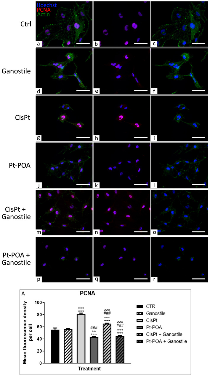
The current standard oncotherapy for glioblastoma is limited by several adverse side effects, leading to a short-term patient survival rate paralleled by a worsening quality of life (QoL). Recently, Complementary and Integrative Medicine's (CIM) innovative approaches have shown positive impacts in terms of better response to treatment, side effect reduction, and QoL improvement. In particular, promising potential in cancer therapy has been found in compounds coming from phyto- and mycotherapy. The objective of this study was to demonstrate the beneficial effects of a new phyto-mycotherapy supplement, named Ganostile, in the human glioblastoma cell line U251, in combination with chemotherapeutic agents, i.e., Cisplatin and a new platinum-based prodrug. Choosing a supplement dosage that mimicked oral supplementation in humans (about 1 g/day), through in vitro assays, microscopy, and cytometric analysis, it has emerged that the cells, after 48hr continuous exposure to Ganostile in combination with the chemical compounds, showed a higher mortality and a lower proliferation rate than the samples subjected to the different treatments administered individually. In conclusion, our data support the use of Ganostile in integrative oncology protocols as a promising adjuvant able to amplify conventional and new drug effects and also reducing resistance mechanisms often observed in brain tumors.

Keywords: drug resistance; ferroptosis; glioblastoma; mitochondrial disfunction; oxidative stress; phytotherapy.


Conflict of interest statement

The authors declare no conflicts of interest.

Figures

Figure 1
Figure 1
Graphic illustration underlining the main results.
Figure 2
Figure 2
MTS viability assay. The cytotoxicity of the Ganostile was evaluated in cultured U251 and primary human fibroblast cells, concentrations from 0.25 to 100 mg/mL were used for 48hr. Cell viability was compared to untreated controls, cultured in the presence of the only basic growing medium. The experiments were performed two times with three technical replicates for each treatment. Data are expressed as mean ± SD referring to the control. p value: (*) < 0.05; (**) < 0.01 and (***) < 0.001.
Figure 3
Figure 3
Cytofluorimetric analysis of DNA content in U251 cells after PI staining in control conditions (CTR) and after treatment with Ganostile at different concentrations: 10 mg/mL, 5 mg/mL, and 1 mg/mL.
Figure 4
Figure 4
Double immunolabeling for PCNA (in red) and actin (in green) in controls (ac) and differently treated U251 cells, i.e., after 48hr-CT using Ganostile 5 mg/mL (df), Cisplatin 40 μM (gi), Pt(IV)Ac-POA 10 μM (jl), Cisplatin + Ganostile (mo), or Pt(IV)Ac-POA + Ganostile (pr). DNA was stained with Hoechst 33258 (blue fluorescence). Magnification 40×, scale bar: 50 μm. (Panel A) Histogram showing the PCNA mean fluorescence intensity per cell (OD) in control and differently treated cells. * control vs. each experimental condition; ° Ganostile vs. other treatments; # Cisplatin vs. any combined treatment; ^ Pt(IV)Ac-POA vs. each combined treatment.
Figure 5
Figure 5
Double immunolabeling for NOS (in green) and actin (in red) in controls (ac) and differently treated U251 cells, i.e., after 48hr-CT using Ganostile 5 mg/mL (df), Cisplatin 40 μM (gi), Pt(IV)Ac-POA 10 μM (jl), Cisplatin + Ganostile (mo), or Pt(IV)Ac-POA + Ganostile (pr). DNA was stained with Hoechst 33258 (blue fluorescence). Magnification 40×; scale bar: 50 μm. (Panel A) Histogram showing the NOS2 mean fluorescence intensity per cell (OD) in control and differently treated cells. * control vs. each experimental condition; ° Ganostile vs. other treatments; # Cisplatin vs. any combined treatment; ^ Pt(IV)Ac-POA vs. each combined treatment; + Cisplatin + Ganostile vs. other experimental groups.
Figure 6
Figure 6
Double immunolabeling for SOD2 (in green) and mitochondria (in red) in controls (ac) and differently treated U251 cells, i.e., after 48hr-CT using Ganostile 5 mg/mL (df), Cisplatin 40 μM (gi), Pt(IV)Ac-POA 10 μM (jl), Cisplatin + Ganostile (mo), or Pt(IV)Ac-POA + Ganostile (pr). DNA was stained with Hoechst 33258 (blue fluorescence). Magnification 40×; scale bar: 50 μm. (Panel A) Histogram showing the SOD2 mean fluorescence intensity per cell (OD) in control and differently treated cells. * control vs. each experimental condition; ° Ganostile vs. other treatments; # Cisplatin vs. any combined treatment; ^ Pt(IV)Ac-POA vs. each combined treatment; + Cisplatin + Ganostile vs. other experimental groups.
Figure 7
Figure 7
Double immunolabeling for COX2 (in red) and mitochondria (in green) in controls (ac) and differently treated U251 cells, i.e., after 48hr-CT using Ganostile 5 mg/mL (df), Cisplatin 40 μM (gi), Pt(IV)Ac-POA 10 μM (jl), Cisplatin + Ganostile (mo), or Pt(IV)Ac-POA + Ganostile (pr). DNA was stained with Hoechst 33258 (blue fluorescence). Magnification 60×; scale bar: 25 μm. (Panel A) Histogram showing the COX2 mean fluorescence intensity per cell (OD) in control and differently treated cells. * control vs. each experimental condition.
Figure 8
Figure 8
Double immunolabeling for COX4 (in green) and mitochondria (in red) in controls (ac) and differently treated U251 cells, i.e., after 48hr-CT using Ganostile 5 mg/mL (df), Cisplatin 40 μM (gi), Pt(IV)Ac-POA 10 μM (jl), Cisplatin + Ganostile (mo), or Pt(IV)Ac-POA + Ganostile (pr). DNA was stained with Hoechst 33258 (blue fluorescence). Magnification 40×; scale bar: 50 μm. (Panel A) Histogram showing the COX4 mean fluorescence intensity per cell (OD) in control and differently treated cells. * control vs. each experimental condition; # Cisplatin vs. any combined treatment; ^ Pt(IV)Ac-POA vs. each combined treatment. ° Ganostile vs. other treatments.
Figure 9
Figure 9
Double immunolabeling for CaM (in green) and actin (in red) in controls (ac) and differently treated U251 cells, i.e., after 48hr-CT using Ganostile 5 mg/mL (df), Cisplatin 40 μM (gi), Pt(IV)Ac-POA 10 μM (jl), Cisplatin + Ganostile (mo), or Pt(IV)Ac-POA + Ganostile (pr). DNA was stained with Hoechst 33258 (blue fluorescence). Magnification 40×; scale bar: 50 μm. (Panel A) Histogram showing the CaM mean fluorescence intensity per cell (OD) in control and differently treated cells.
Figure 10
Figure 10
Double immunolabeling for caspase-3 (in green) and actin (in red) in controls (ac) and differently treated U251 cells, i.e., after 48hr-CT using Ganostile 5 mg/mL (df), Cisplatin 40 μM (gi), Pt(IV)Ac-POA 10 μM (jl), Cisplatin + Ganostile (mo), or Pt(IV)Ac-POA + Ganostile (pr). DNA was stained with Hoechst 33258 (blue fluorescence). Magnification 40×; scale bar: 50 μm. (Panel A) Histogram showing the caspase-3 mean fluorescence intensity per cell (OD) in control and differently treated cells. * control vs. each experimental condition; ° Ganostile vs. other treatments; # Cisplatin vs. any combined treatment; ^ Pt(IV)Ac-POA vs. each combined treatment.
Figure 11
Figure 11
Double immunolabeling for GPX4 (in green) and actin (in red) in controls (ac) and differently treated U251 cells, i.e., after 48hr-CT using Ganostile 5 mg/mL (df), Cisplatin 40 μM (gi), Pt(IV)Ac-POA 10 μM (jl), Cisplatin + Ganostile (mo), or Pt(IV)Ac-POA + Ganostile (pr). DNA was stained with Hoechst 33258 (blue fluorescence). Magnification 40×; scale bar: 50 μm. (Panel A) Histogram showing the GPX4 mean fluorescence intensity per cell (OD) in control and differently treated cells. * control vs. each experimental condition; ° Ganostile vs. other treatments; # Cisplatin vs. any combined treatment; ^ Pt(IV)Ac-POA vs. each combined treatment.
Figure 12
Figure 12
Double immunolabeling for LC3b (in green) and lysosomes (in red) in controls (ac) and differently treated U251 cells, i.e., after 48hr-CT using Ganostile 5 mg/mL (df), Cisplatin 40 μM (gi), Pt(IV)Ac-POA 10 μM (jl), Cisplatin + Ganostile (mo), or Pt(IV)Ac-POA + Ganostile (pr). DNA was stained with Hoechst 33258 (blue fluorescence). Magnification 60×; scale bar: 25 μm. (Panel A) Histogram showing the LC3b mean fluorescence intensity per cell (OD) in control and differently treated cells. * control vs. each experimental condition; ° Ganostile vs. other treatments; # Cisplatin vs. any combined treatment; ^ Pt(IV)Ac-POA vs. each combined treatment; + Cisplatin + Ganostile vs. other experimental groups.
Figure 13
Figure 13
Double immunolabeling for p62 (in green) and lysosomes (in red) in controls (ac) and differently treated U251 cells, i.e., after 48hr-CT using Ganostile 5 mg/mL (df), Cisplatin 40 μM (gi), Pt(IV)Ac-POA 10 μM (jl), Cisplatin + Ganostile (mo), or Pt(IV)Ac-POA + Ganostile (pr). DNA was stained with Hoechst 33258 (blue fluorescence). Magnification 60×; scale bar: 25 μm. (Panel A) Histogram showing the p62 mean fluorescence intensity per cell (OD) in control and differently treated cells. * control vs. each experimental condition; ° Ganostile vs. other treatments; ^ Pt(IV)Ac-POA vs. each combined treatment; + Cisplatin + Ganostile vs. other experimental groups.
Figure 14
Figure 14
Double immunolabeling for PINK1 (in green) and mitochondria (in red) in controls (ac) and differently treated U251 cells, i.e., after 48hr-CT using Ganostile 5 mg/mL (df), Cisplatin 40 μM (gi), Pt(IV)Ac-POA 10 μM (jl), Cisplatin + Ganostile (mo), or Pt(IV)Ac-POA + Ganostile (pr). DNA was stained with Hoechst 33258 (blue fluorescence). Magnification 40×; scale bar: 50 μm. (Panel A) Histogram showing the PINK1 mean fluorescence intensity per cell (OD) in control and differently treated cells. * control vs. each experimental condition; ° Ganostile vs. other treatments; # Cisplatin vs. any combined treatment.
Figure 15
Figure 15
Double immunolabeling for Parkin (in green) and mitochondria (in red) in controls (ac) and differently treated U251 cells, i.e., after 48hr-CT using Ganostile 5 mg/mL (df), Cisplatin 40 μM (gi), Pt(IV)Ac-POA 10 μM (jl), Cisplatin + Ganostile (mo), or Pt(IV)Ac-POA + Ganostile (pr). DNA was stained with Hoechst 33258 (blue fluorescence). Magnification 40×; scale bar: 50 μm. (Panel A) Histogram showing the Parkin mean fluorescence intensity per cell (OD) in control and differently treated cells. * control vs. each experimental condition; ° Ganostile vs. other treatments; # Cisplatin vs. any combined treatment; ^ Pt(IV)Ac-POA vs. each combined treatment; + Cisplatin + Ganostile vs. other experimental groups.
Figure 16
Figure 16
Immunolabeling for actin (a,e,i,m,q, and u, in red), mitochondria (b,f,j,n,r, and v, in green), lysosomes (c,g,k,o,s, and w, in red) and Golgi apparatus (d,h,l,p,t, and x, in red) in controls (ad) and differently treated U251 cells, i.e., after 48hr-CT using Ganostile 5 mg/mL (eh), Cisplatin 40 μM (il), Pt(IV)Ac-POA 10 μM (mp), Cisplatin + Ganostile (qt), or Pt(IV)Ac-POA + Ganostile (ux). DNA was stained with Hoechst 33258 (blue fluorescence). Magnification 40×; scale bar: 50 μm (a,d,e,h,i,l,m,p,q,t,u, and x); 60×; scale bar: 25 μm (b,c,f,g,j,k,n,o,r,s,v, and w).
Figure 17
Figure 17
Histogram showing the (Panel A) actin, (Panel B) mitochondria, (Panel C) lysosomes, and (Panel D) Golgi apparatus mean fluorescence intensity per cell (OD) in control and differently treated cells. * control vs. each experimental condition; ° Ganostile vs. other treatments; # Cisplatin vs. any combined treatment; ^ Pt(IV)Ac-POA vs. each combined treatment.
Figure 18
Figure 18
TEM ultrastructural analysis of control (a) and Ganostile 5 mg/mL (b), Cisplatin 40 μM (c), Cisplatin + Ganostile (d), Pt(IV)Ac-POA 10 μM (e), and Pt(IV)Ac-POA + Ganostile (f) treated cells. N: nucleus. Scale bar; 2 μm (a,cf); 5 μm (b).
Figure 19
Figure 19
Chemical structure of Pt(IV)Ac-POA.

Similar articles

Cited by

References

    1. Stupp R., Mason W.P., van den Bent M.J., Weller M., Fisher B., Taphoorn M.J.B., Belanger K., Brandes A.A., Marosi C., Bogdahn U., et al. Radiotherapy plus Concomitant and Adjuvant Temozolomide for Glioblastoma. N. Engl. J. Med. 2005;352:987–996. doi: 10.1056/nejmoa043330. - DOI - PubMed
    1. Rossi P., Difrancia R., Quagliariello V., Savino E., Tralongo P., Randazzo C.L., Berretta M. B-Glucans from Grifola Frondosa and Ganoderma Lucidum in Breast Cancer: An Example of Complementary and Integrative Medicine. Oncotarget. 2018;9:24837–24856. doi: 10.18632/oncotarget.24984. - DOI - PMC - PubMed
    1. Qu L., Li S., Zhuo Y., Chen J., Qin X., Guo G. Anticancer Effect of Triterpenes from Ganoderma Lucidum in Human Prostate Cancer Cells. Oncol. Lett. 2017;14:7467–7472. doi: 10.3892/ol.2017.7153. - DOI - PMC - PubMed
    1. Jiang D., Wang L., Zhao T., Zhang Z., Zhang R., Jin J., Cai Y., Wang F. Restoration of the Tumor-Suppressor Function to Mutant P53 by Ganoderma Lucidum Polysaccharides in Colorectal Cancer Cells. Oncol. Rep. 2017;37:594–600. doi: 10.3892/or.2016.5246. - DOI - PubMed
    1. Ferreira I.C.F.R., Heleno S.A., Reis F.S., Stojkovic D., Queiroz M.J.R.P., Vasconcelos M.H., Sokovic M. Chemical Features of Ganoderma Polysaccharides with Antioxidant, Antitumor and Antimicrobial Activities. Phytochemistry. 2015;114:38–55. doi: 10.1016/j.phytochem.2014.10.011. - DOI - PubMed

MeSH terms