. 2024 Jun 5;25(11):6211.
doi: 10.3390/ijms25116211.

Galanin Coordinates Macrophage-Associated Fibro-Inflammatory Response and Mitochondrial Integrity in Myocardial Infarction Reperfusion Injury

Affiliations

Galanin Coordinates Macrophage-Associated Fibro-Inflammatory Response and Mitochondrial Integrity in Myocardial Infarction Reperfusion Injury

Lesia Savchenko et al. Int J Mol Sci. .

Abstract

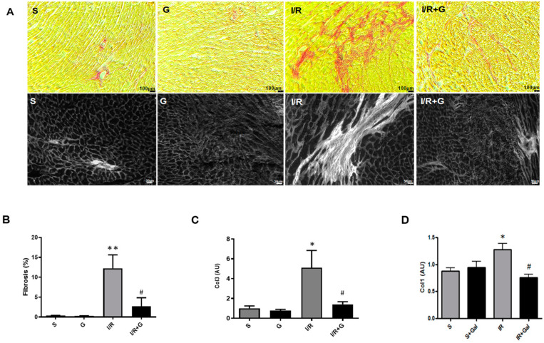
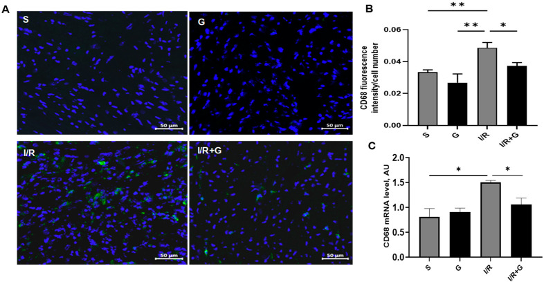
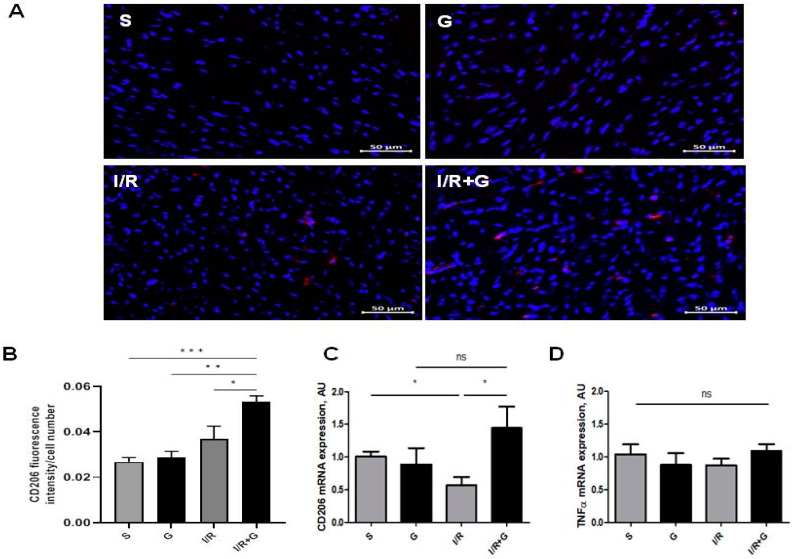
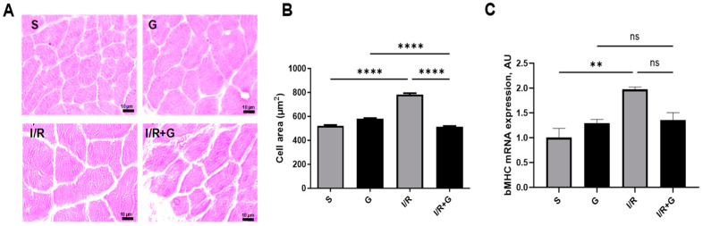
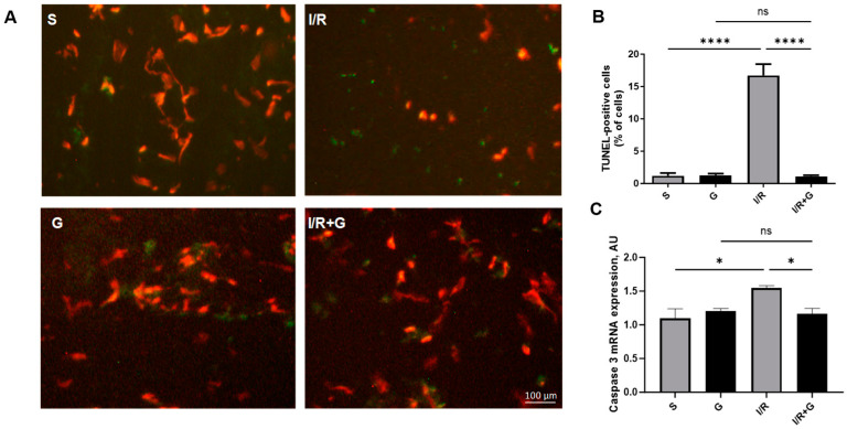
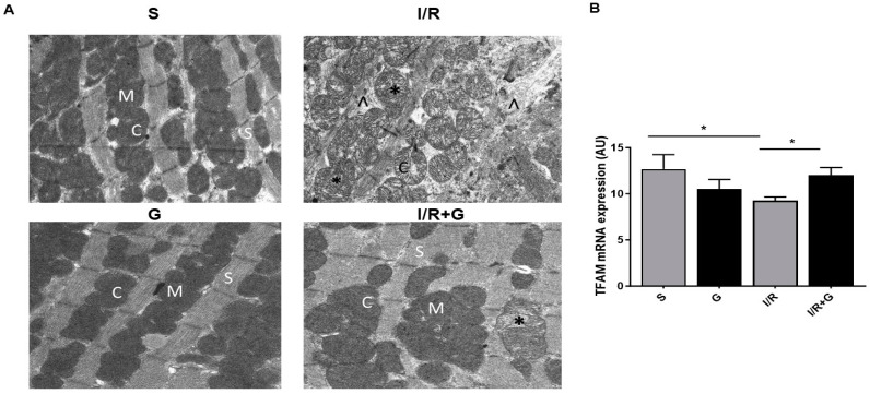
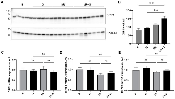
Myocardial infarction activates an intense fibro-inflammatory reaction that is essential for cardiac remodeling and heart failure (HF). Bioactive peptide galanin plays a critical role in regulating cardiovascular homeostasis; however, its specific functional relevance in post-infarction fibro-inflammatory reprogramming remains obscure. Here, we show that galanin coordinates the fibro-inflammatory trajectory and mitochondrial integrity in post-infarction reperfusion injury. Aberrant deposition of collagen was associated with a marked increase in CD68-positive macrophage infiltration in cardiac tissue in mice subjected to myocardial ischemia/reperfusion (I/R) for 14 days compared to sham controls. Furthermore, we found that the myocardial expression level of a specific marker of M2 macrophages, CD206, was significantly down-regulated in I/R-challenged mice. In contrast, galanin treatment started during the reperfusion phase blunted the fibro-inflammatory responses and promoted the expression of CD206 in I/R-remodeled hearts. In addition, we found that the anti-apoptotic and anti-hypertrophic effects of galanin were associated with the preservation of mitochondrial integrity and promotion of mitochondrial biogenesis. These findings depict galanin as a key arbitrator of fibro-inflammatory responses to cardiac I/R injury and offer a promising therapeutic trajectory for the treatment of post-infarct cardiovascular complications.

Keywords: apoptosis; cardiac remodeling; fibrosis; inflammation; mitochondria.


Conflict of interest statement

The authors declare no conflicts of interest.

Figures

Figure 1
Figure 1
Galanin blunts cardiac fibrosis in myocardial infarction reperfusion injury. (A) Representative images of cardiac sections stained with Sirius Red (top panel; collagen fibers: red color) and visualized by polarized light microscopy (bottom panel; collagen fibers: white color) from mice subjected to sham operation or 30 min of cardiac ischemia and 14 days of reperfusion (I/R). (B) Quantification of myocardial fibrosis in (A). (C,D) Myocardial mRNA levels of collagen 1 (Col 1) and collagen 3 (Col 3) in vehicle- or galanin-treated (G) mice after 14 days of I/R injury, n = 5. Data presented as the mean ± SEM. * p < 0.05, ** p < 0.01 vs. sham, # p < 0.05 vs. I/R according to one-way ANOVA followed by Bonferroni’s post hoc test.
Figure 2
Figure 2
Galanin inhibits macrophage-associated inflammatory reactions in myocardial infarction reperfusion injury. (A) CD68 immunostaining (green fluorescence) was performed to detect inflammatory cell populations in cardiac sections from vehicle- or galanin-treated mice subjected to 30 min of cardiac ischemia and 14 days of reperfusion. Nuclei were counterstained with DAPI (blue). (B) Quantification of CD68-positive cells from (A), n = 5. (C) Myocardial CD68 mRNA levels in vehicle- or galanin-treated mice subjected to I/R or sham, n = 5. Data presented as the mean ± SEM. * p < 0.05, ** p < 0.01 according to one-way ANOVA followed by Bonferroni’s post hoc test.
Figure 3
Figure 3
Galanin promotes the anti-inflammatory trajectory in I/R-remodeled hearts. Mice were exposed to 30 min of cardiac ischemia and 14 days of reperfusion. (A,B) Representative images and quantification of CD206 (red) in vehicle- or galanin-treated mice subjected to I/R damage, n = 4. Nuclei were counterstained with DAPI (blue). (C,D) Myocardial mRNA levels of CD206 and TNFα in vehicle- or galanin-treated control sham and I/R-challenged mice, n = 5. Data presented as the mean ± SEM; ns: not significant. * p < 0.05, ** p < 0.01, *** p < 0.001, according to one-way ANOVA followed by Bonferroni’s post hoc test.
Figure 4
Figure 4
Galanin reduces cardiac hypertrophy in myocardial infarction reperfusion injury. (A) Representative photomicrographs of cardiac tissue sections with H&E staining and (B) quantitative analysis of the cross-sectional area. (C) Myocardial mRNA levels of β-MHC in vehicle- or galanin-treated control sham and I/R-challenged mice, n = 5. Data presented as the mean ± SEM; ns: not significant. ** p < 0.01, **** p < 0.001 according to one-way ANOVA followed by Bonferroni’s post hoc test.
Figure 5
Figure 5
Galanin prevents myocardial apoptosis in I/R-challenged hearts. (A) Representative photomicrographs of TUNEL staining of cardiac sections from vehicle- or galanin-treated mice exposed to 30 min of cardiac ischemia and 14 days of reperfusion. The green cells indicate the TUNEL-positive apoptotic cells. (B) Quantification of TUNEL-positive cells from (A), n = 5. (C) Myocardial mRNA levels (C) of caspase 3 in vehicle- or galanin-treated mice exposed to I/R injury. Data presented as the mean ± SEM; ns: not significant. * p < 0.05, **** p < 0.0001 according to one-way ANOVA followed by Bonferroni’s post hoc test.
Figure 6
Figure 6
Galanin preserves mitochondrial integrity in myocardial infarction reperfusion injury. (A) Electron micrographs of hearts from vehicle- or galanin (G)-treated mice subjected to cardiac I/R injury. Cardiac tissues were harvested for examination of the myocardial ultrastructure by transmission electron microscopy. Magnification: 14,000×. (M)—mitochondria; (C)—cristae; (S)—sarcomere; (*)—damaged mitochondria; (∧)—disrupted myofibrils. (B) Myocardial TFAM mRNA levels in vehicle- or galanin-treated mice subjected to I/R or sham. n = 5. Data are expressed as the mean ± SEM. * p < 0.05 according to one-way ANOVA followed by Bonferroni’s post hoc test.
Figure 7
Figure 7
Galanin promotes mitochondrial fission in I/R-remodeled hearts. (A) Representative Western blot of DRP1 protein expression and (B) quantification of (A). Myocardial mRNA levels of (C) DRP1, (D) MFN1, and (E) MFN2 in vehicle- or galanin-treated control sham and I/R-challenged mice, n = 5. Mice were exposed to 30 min of cardiac ischemia and 14 days of reperfusion. Data presented as the mean ± SEM; ns: not significant. ** p < 0.01 according to one-way ANOVA followed by Bonferroni’s post hoc test.

Similar articles

Cited by

References

    1. Liu Y., Li L., Wang Z., Zhang J., Zhou Z. Myocardial ischemia-reperfusion injury; Molecular mechanisms and prevention. Microvasc. Res. 2023;149:104565. doi: 10.1016/j.mvr.2023.104565. - DOI - PubMed
    1. Yin X., Yin X., Pan X., Zhang J., Fan X., Li J., Zhai X., Jiang L., Hao P., Wang J., et al. Post-myocardial infarction fibrosis: Pathophysiology, examination, and intervention. Front. Pharmacol. 2023;14:1070973. doi: 10.3389/fphar.2023.1070973. - DOI - PMC - PubMed
    1. de Oliveira Camargo R., Abual’Anaz B., Rattan S.G., Filomeno K.L., Dixon I.M.C. Novel factors that activate and deactivate cardiac fibroblasts: A new perspective for treatment of cardiac fibrosis. Wound Repair Regen. 2021;29:667–677. doi: 10.1111/wrr.12947. - DOI - PubMed
    1. Osokina A., Karetnikova V., Polikutina O., Ivanova A., Gruzdeva O., Dyleva Y., Kokov A., Brel N., Pecherina T., Barbarash O. Prognostic potential of cardiac structural and functional parameters and N-terminal propeptide of type III procollagen in predicting cardiac fibrosis one year after myocardial infarction with preserved left ventricular ejection fraction. Aging. 2021;13:194–203. doi: 10.18632/aging.202495. - DOI - PMC - PubMed
    1. Ren Z., Yang K., Zhao M., Liu W., Zhang X., Chi J., Shi Z., Zhang X., Fu Y., Liu Y., et al. Calcium-Sensing Receptor on Neutrophil Promotes Myocardial Apoptosis and Fibrosis after Acute Myocardial Infarction via NLRP3 in Flammasome Activation. Can. J. Cardiol. 2020;36:893–905. doi: 10.1016/j.cjca.2019.09.026. - DOI - PubMed

MeSH terms