. 2024 Jun 5;25(11):6212.
doi: 10.3390/ijms25116212.

Does Cell-Type-Specific Silencing of Monoamine Oxidase B Interfere with the Development of Right Ventricle (RV) Hypertrophy or Right Ventricle Failure in Pulmonary Hypertension?

Affiliations

Does Cell-Type-Specific Silencing of Monoamine Oxidase B Interfere with the Development of Right Ventricle (RV) Hypertrophy or Right Ventricle Failure in Pulmonary Hypertension?

Paulin Brosinsky et al. Int J Mol Sci. .

Abstract

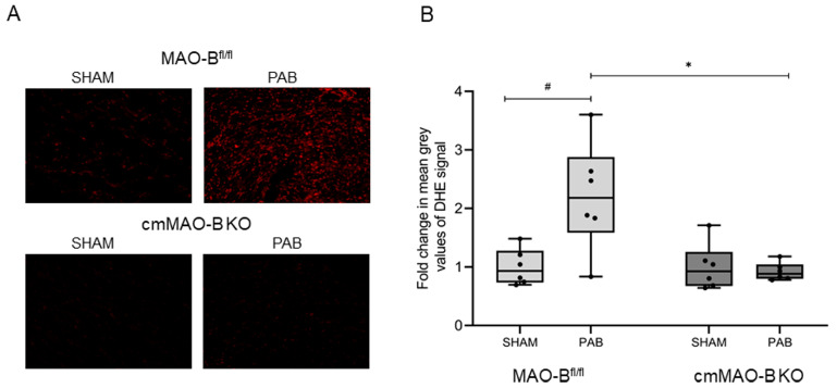
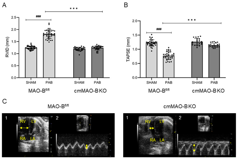
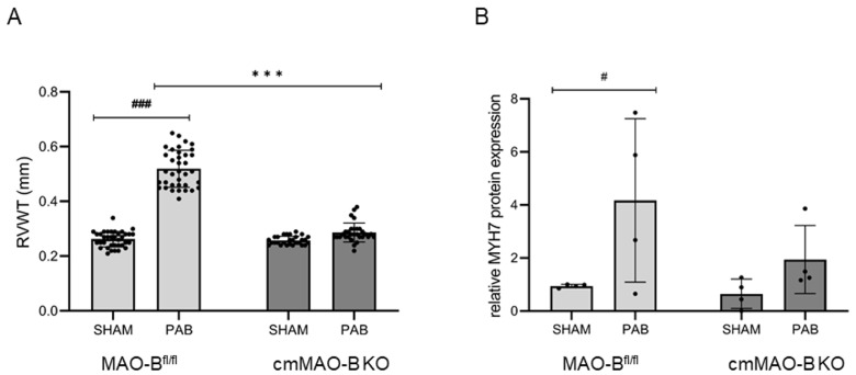
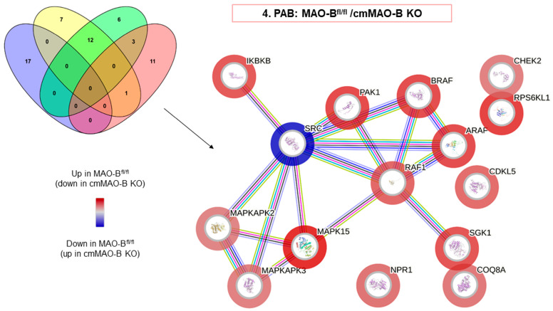
Increased mitochondrial reactive oxygen species (ROS) formation is important for the development of right ventricular (RV) hypertrophy (RVH) and failure (RVF) during pulmonary hypertension (PH). ROS molecules are produced in different compartments within the cell, with mitochondria known to produce the strongest ROS signal. Among ROS-forming mitochondrial proteins, outer-mitochondrial-membrane-located monoamine oxidases (MAOs, type A or B) are capable of degrading neurotransmitters, thereby producing large amounts of ROS. In mice, MAO-B is the dominant isoform, which is present in almost all cell types within the heart. We analyzed the effect of an inducible cardiomyocyte-specific knockout of MAO-B (cmMAO-B KO) for the development of RVH and RVF in mice. Right ventricular hypertrophy was induced by pulmonary artery banding (PAB). RV dimensions and function were measured through echocardiography. ROS production (dihydroethidium staining), protein kinase activity (PamStation device), and systemic hemodynamics (in vivo catheterization) were assessed. A significant decrease in ROS formation was measured in cmMAO-B KO mice during PAB compared to Cre-negative littermates, which was associated with reduced activity of protein kinases involved in hypertrophic growth. In contrast to littermates in which the RV was dilated and hypertrophied following PAB, RV dimensions were unaffected in response to PAB in cmMAO-B KO mice, and no decline in RV systolic function otherwise seen in littermates during PAB was measured in cmMAO-B KO mice. In conclusion, cmMAO-B KO mice are protected against RV dilatation, hypertrophy, and dysfunction following RV pressure overload compared to littermates. These results support the hypothesis that cmMAO-B is a key player in causing RV hypertrophy and failure during PH.

Keywords: monoamine oxidase; pulmonary hypertension; right heart.


Conflict of interest statement

The authors declare no conflicts of interest.

Figures

Figure 1
Figure 1
Increased ROS production in response to PAB was abrogated in cmMAO-B KO mice. Representative images (20× microscope’s magnification) of cryosections of RV tissue stained with dihydroethidium (DHE) dye from PAB- or SHAM-operated MAO-Bfl/fl and cmMAO-B KO mice (A). Quantification of ROS formation in RV tissue from MAO-Bfl/fl (SHAM and PAB n = 6 each) and cmMAO-B KO (SHAM and PAB n = 6 each) mice by measuring mean grey values of the DHE signal. Data are presented as mean fold change compared to MAO-Bfl/fl SHAM ± SD, * and #: p < 0.05 analyzed through two-side ANOVA (B).
Figure 2
Figure 2
PAB-operated cmMAO-B KO mice were unaffected in their RV dimension and systolic function. RV geometry and function of MAO-Bfl/fl (SHAM n = 33, PAB n = 33) and cmMAO-B KO (SHAM n = 22, PAB n = 23) mice were measured through echocardiography three weeks after PAB or SHAM surgery. Data are shown for right ventricular inner diameter (RVID in mm) (A) and tricuspid annular plane systolic excursion (TAPSE in mm) (B). The dots represent the individual data points. Data represent the mean ± SD, ### and ***: p < 0.005 analyzed through two-side ANOVA. Images of apical four-chamber view (1) and the systolic excursion of the tricuspid valve (TAPSE) (2) of one representative MAO-Bfl/fl and cmMAO-B KO heart (C). LA: left atrium, LV: left ventricle, RA: right atrium, RV: right ventricle. Horizontal arrows represent RVID; vertical arrows represent TAPSE.
Figure 3
Figure 3
Cardiomyocyte-specific deletion of MAO-B protected the hearts from RVH. Three weeks after SHAM/PAB surgery, mice were evaluated for RVH development. Echocardiography was used to determine right ventricular wall thickness RVWT (in mm). Each dot represents the individual data point. Data represent the mean ± SD for MAO-Bfl/fl (SHAM n = 36, PAB n = 36) and cmMAO-B KO mice (SHAM n = 28, PAB n = 30) (A). The Jess Simple Western system was used to detect the hypertrophy marker Myosin Heavy Chain (MYH) 7 at the protein level in the right part of the septum of MAO-Bfl/fl and cmMAO-B KO mice (n = 4 for each of the four conditions) normalized to vinculin. Data represent the mean ± SD relative to the MAO-Bfl/fl SHAM group (B). Both diagrams were analyzed through two-side ANOVA, #: p < 0.05, ### and ***: p < 0.005.
Figure 4
Figure 4
The activity of the kinases involved in cardiac hypertrophy was decreased in cmMAO-B KO mice in response to pressure overload. Functional protein association networks of kinases were analyzed in the right part of the septum of mice due pressure overload. MAO-Bfl/fl and cmMAO-B KO mice underwent PAB surgery (n = 4, for each group). After three weeks, protein lysates from the right part of the septum were prepared for kinome profiling. Sequential two-group comparisons were performed to identify kinases, which are differentially regulated because of the genetic background and in response to pressure overload. The resulting hits were uploaded into the String protein database webpage to create images representing functional relationships revealing protein, i.e., kinase, association networks. The Venn diagram highlights the number of kinases that are significantly (p < 0.05) de-regulated in not only one but multiple two-group comparisons. For the reactome diagram, the outer color reflects the kinase activity highlighted for PAB: MAO-Bfl/fl vs. cmMAO-B KO.

Similar articles

Cited by

References

    1. Maarman G.J., Schulz R., Sliwa K., Schermuly R.T., Lecour S. Novel putative pharmacological therapies to protect the right ventricle in pulmonary hypertension: A review of current literature. Br. J. Pharmacol. 2017;174:497–511. doi: 10.1111/bph.13721. - DOI - PMC - PubMed
    1. Schlüter K.-D., Kutsche H.S., Hirschhäuser C., Schreckenberg R., Schulz R. Review on Chamber-Specific Differences in Right and Left Heart Reactive Oxygen Species Handling. Front. Physiol. 2018;9:1799. doi: 10.3389/fphys.2018.01799. - DOI - PMC - PubMed
    1. Schulz R., Schlüter K.-D. Importance of Mitochondria in Cardiac Pathologies: Focus on Uncoupling Proteins and Monoamine Oxidases. Int. J. Mol. Sci. 2023;24:6459. doi: 10.3390/ijms24076459. - DOI - PMC - PubMed
    1. Heusch P., Canton M., Aker S., Van De Sand A., Konietzka I., Rassaf T., Menazza S., Brodde O., Di Lisa F., Heusch G., et al. The contribution of reactive oxygen species and p38 mitogen-activated protein kinase to myofilament oxidation and progression of heart failure in rabbits. Br. J. Pharmacol. 2010;160:1408–1416. doi: 10.1111/j.1476-5381.2010.00793.x. - DOI - PMC - PubMed
    1. Andreadou I., Schulz R., Papapetropoulos A., Turan B., Ytrehus K., Ferdinandy P., Daiber A., Di Lisa F. The role of mitochondrial reactive oxygen species, NO and H 2 S in ischaemia/reperfusion injury and cardioprotection. J. Cell. Mol. Med. 2020;24:6510–6522. doi: 10.1111/jcmm.15279. - DOI - PMC - PubMed

MeSH terms