. 2024 Jun 6;25(11):6257.
doi: 10.3390/ijms25116257.

Exploring the Ocular Surface Microbiome and Tear Proteome in Glaucoma

Affiliations

Exploring the Ocular Surface Microbiome and Tear Proteome in Glaucoma

Livia Spörri et al. Int J Mol Sci. .

Abstract

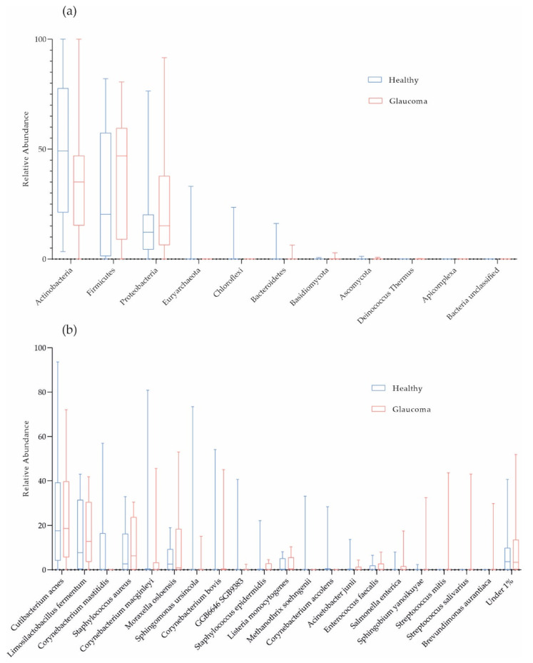
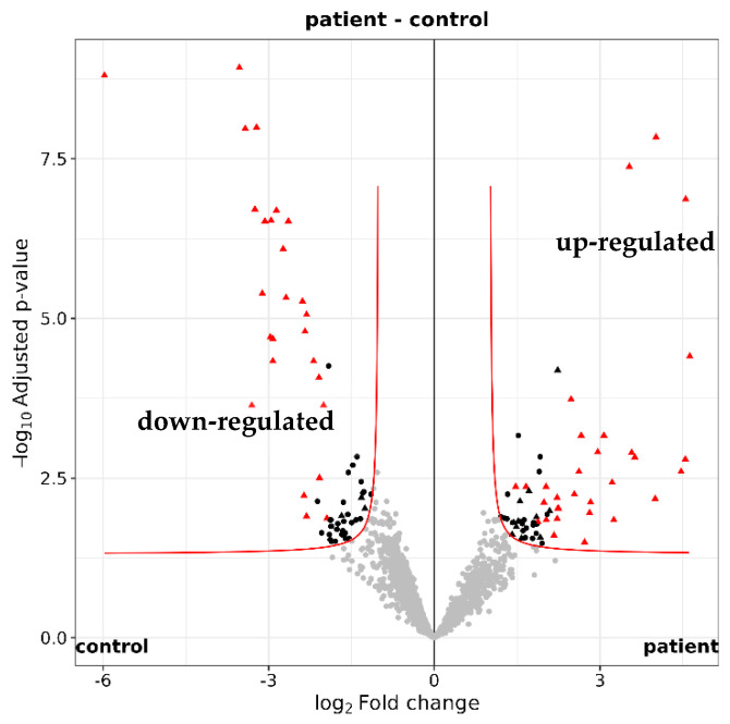
Although glaucoma is a leading cause of irreversible blindness worldwide, its pathogenesis is incompletely understood, and intraocular pressure (IOP) is the only modifiable risk factor to target the disease. Several associations between the gut microbiome and glaucoma, including the IOP, have been suggested. There is growing evidence that interactions between microbes on the ocular surface, termed the ocular surface microbiome (OSM), and tear proteins, collectively called the tear proteome, may also play a role in ocular diseases such as glaucoma. This study aimed to find characteristic features of the OSM and tear proteins in patients with glaucoma. The whole-metagenome shotgun sequencing of 32 conjunctival swabs identified Actinobacteria, Firmicutes, and Proteobacteria as the dominant phyla in the cohort. The species Corynebacterium mastitidis was only found in healthy controls, and their conjunctival microbiomes may be enriched in genes of the phospholipase pathway compared to glaucoma patients. Despite these minor differences in the OSM, patients showed an enrichment of many tear proteins associated with the immune system compared to controls. In contrast to the OSM, this emphasizes the role of the proteome, with a potential involvement of immunological processes in glaucoma. These findings may contribute to the design of new therapeutic approaches targeting glaucoma and other associated diseases.

Keywords: glaucoma; liquid chromatography–tandem mass spectrometry; ocular surface microbiome; tear proteome; whole-metagenome shotgun sequencing.


Conflict of interest statement

The authors declare no conflicts of interest. The funders had no role in the design of the study; in the collection, analyses, or interpretation of data; in the writing of the manuscript, or in the decision to publish the results. Throughout the entirety of the work, ChatGPT 3.5 was utilized for structural assistance in writing the paper, as well as for linguistic and formal revisions.

Figures

Figure 1
Figure 1
Taxonomic characterization of ocular surface microbiome. Box plots for relative abundances at phylum (a) and species level (b) are shown (red, glaucoma, n = 16; blue, healthy controls, n = 16).
Figure 2
Figure 2
Diversity in the ocular surface microbiome. There were no differences in Shannon index observed between glaucoma patients (n = 16) and controls (n = 16; p = 0.35, Welch’s t test).
Figure 3
Figure 3
Principal component analysis (PCA) of microbial abundances (a) and protein intensities (b). (a) PCA of microbial abundances did not separate glaucoma patients (red, n = 16) from healthy controls (blue, n = 16; p = 0.51, PERMANOVA, n repeat = 1000). (b) Log2−transformed protein group intensities before imputation were used for analysis. PCA separates glaucoma patients (n = 16) from controls (n = 16; p = 0.0060, PERMANOVA, n repeat = 1000). Orange, female glaucoma patients (n = 7); red, male glaucoma patients (n = 9); light blue, healthy control females (n = 10); blue, healthy control males (n = 6).
Figure 4
Figure 4
The ocular surface microbiome of POAG and PEXG patients. Box plots for relative abundances of all species are shown. PEXG, pseudoexfoliation glaucoma (n = 8); POAG, primary open-angle glaucoma (n = 8).
Figure 5
Figure 5
Quantification of the tear proteome. Volcano plot illustrating the 2250 identifed tear proteins. A total of 123 proteins with a maximum adjusted p value of 0.05 were either up-regulated (log2 fold change ≥ 1) or down-regulated (log2 fold change ≤ −1) in glaucoma patients (n = 16) versus healthy controls (n = 16).

Similar articles

Cited by

References

    1. Weinreb R.N., Aung T., Medeiros F.A. The pathophysiology and treatment of glaucoma: A review. JAMA. 2014;311:1901–1911. doi: 10.1001/jama.2014.3192. - DOI - PMC - PubMed
    1. Agarwal R., Gupta S.K., Agarwal P., Saxena R., Agrawal S.S. Current concepts in the pathophysiology of glaucoma. Indian J. Ophthalmol. 2009;57:257–266. - PMC - PubMed
    1. Shon K., Wollstein G., Schuman J.S., Sung K.R. Prediction of glaucomatous visual field progression: Pointwise analysis. Curr. Eye Res. 2014;39:705–710. doi: 10.3109/02713683.2013.867353. - DOI - PMC - PubMed
    1. Tham Y.C., Li X., Wong T.Y., Quigley H.A., Aung T., Cheng C.Y. Global prevalence of glaucoma and projections of glaucoma burden through 2040: A systematic review and meta-analysis. Ophthalmology. 2014;121:2081–2090. doi: 10.1016/j.ophtha.2014.05.013. - DOI - PubMed
    1. Huang L., Hong Y., Fu X., Tan H., Chen Y., Wang Y., Chen D. The role of the microbiota in glaucoma. Mol. Asp. Med. 2023;94:101221. doi: 10.1016/j.mam.2023.101221. - DOI - PubMed