. 2024 Jun 6;25(11):6279.
doi: 10.3390/ijms25116279.

EGFRvIII Confers Sensitivity to Saracatinib in a STAT5-Dependent Manner in Glioblastoma

Affiliations

EGFRvIII Confers Sensitivity to Saracatinib in a STAT5-Dependent Manner in Glioblastoma

Mylan R Blomquist et al. Int J Mol Sci. .

Abstract

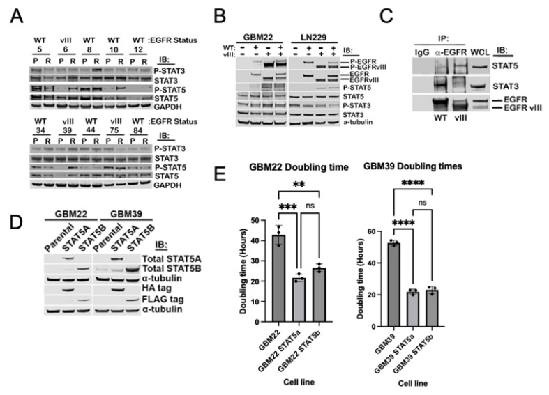
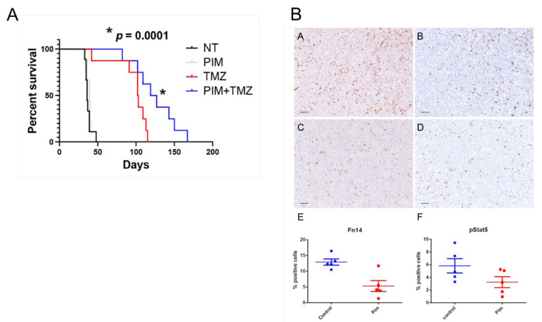
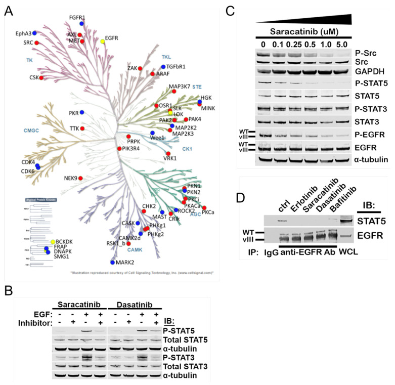
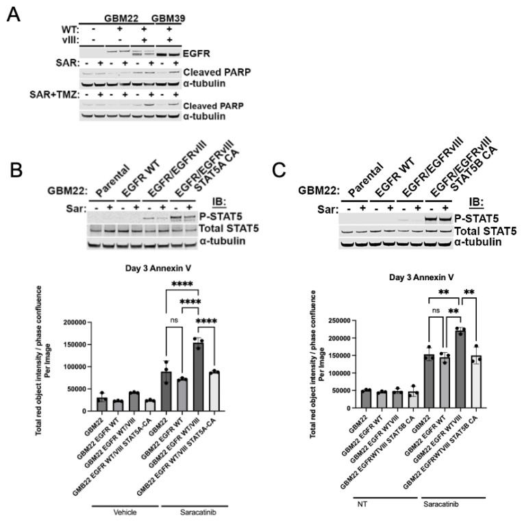
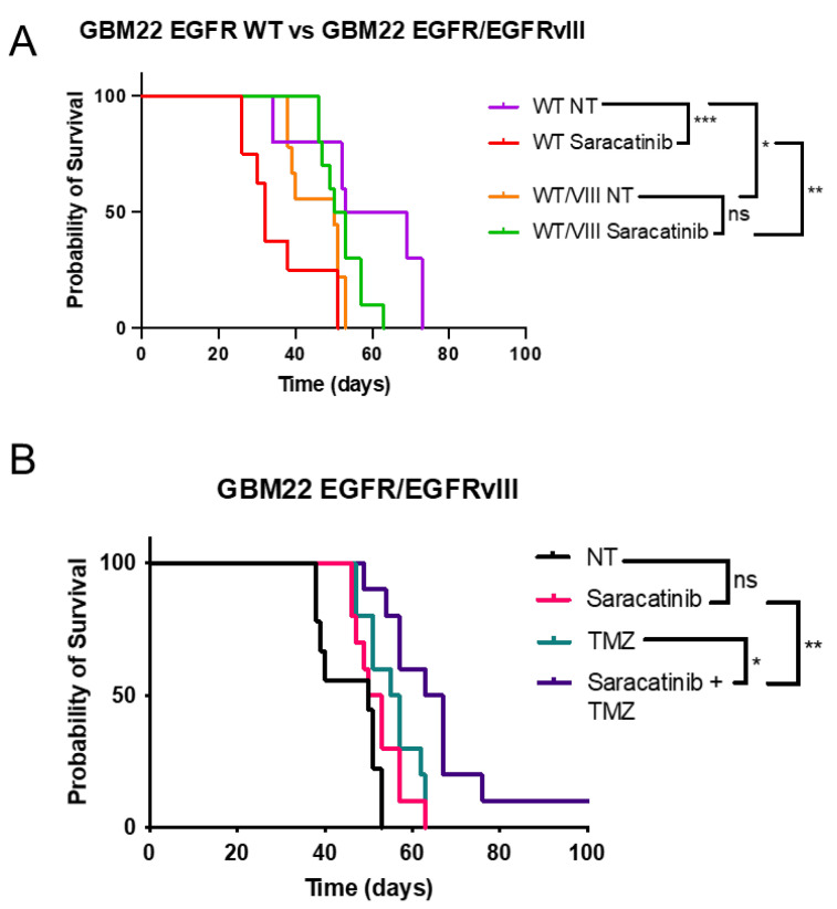
Glioblastoma (GBM) is the most common primary malignant brain tumor in adults, with few effective treatments. EGFR alterations, including expression of the truncated variant EGFRvIII, are among the most frequent genomic changes in these tumors. EGFRvIII is known to preferentially signal through STAT5 for oncogenic activation in GBM, yet targeting EGFRvIII has yielded limited clinical success to date. In this study, we employed patient-derived xenograft (PDX) models expressing EGFRvIII to determine the key points of therapeutic vulnerability within the EGFRvIII-STAT5 signaling axis in GBM. Our findings reveal that exogenous expression of paralogs STAT5A and STAT5B augments cell proliferation and that inhibition of STAT5 phosphorylation in vivo improves overall survival in combination with temozolomide (TMZ). STAT5 phosphorylation is independent of JAK1 and JAK2 signaling, instead requiring Src family kinase (SFK) activity. Saracatinib, an SFK inhibitor, attenuates phosphorylation of STAT5 and preferentially sensitizes EGFRvIII+ GBM cells to undergo apoptotic cell death relative to wild-type EGFR. Constitutively active STAT5A or STAT5B mitigates saracatinib sensitivity in EGFRvIII+ cells. In vivo, saracatinib treatment decreased survival in mice bearing EGFR WT tumors compared to the control, yet in EGFRvIII+ tumors, treatment with saracatinib in combination with TMZ preferentially improves survival.

Keywords: cell signaling; glioblastoma; precision oncology; receptor tyrosine kinase.


Conflict of interest statement

Author Landon Inge was employed by the company Ventana Medical Systems, Roche Diagnostics. The remaining authors declare that the research was conducted in the absence of any commercial or financial relationships that could be construed as a potential conflict of interest.

Figures

Figure 1
Figure 1
STAT5, active downstream of EGFRvIII in GBM, promotes cell proliferation in vitro. (A) Lysates from patient-derived xenograft tissues, either the parental tumor (P) or the induced temozolomide resistant tissue (R), were immunoblotted for phosphorylated and total STAT3 and STAT5. EGFR status is indicated above patient-derived xenograft number. (B) STAT5 is only phosphorylated downstream of EGFRvIII in GBM. Cell lines LN229 and GBM22 stably expressing either EGFR WT, EGFRvIII, or both constructs (EGFR/EGFRvIII) were serum starved, lysed, and immunoblotted for the indicated antibodies. (C) GBM22 expressing either EGFR WT or EGFRvIII was lysed and immunoprecipitated with an anti−EGFR antibody against the cytoplasmic domain (Abcam 52894; Abcam, Cambridge, UK). Precipitates were immunoblotted for total STAT5 and total STAT3. WCL whole cell lysate. (D) GBM22 and GBM39 stably expressing STAT5A or STAT5B were serum starved, lysed, and probed for both STAT5A/B expression and the appropriate protein tag. (E) Doubling time for GBM22 and GBM39 stably expressing STAT5A or STAT5B was calculated based on the average of initial cell confluence and the average of confluence at 60 h from three individual experiments measured by Incucyte SX5. **** p < 0.0001, *** p < 0.0005, ** p < 0.005, ns = not significant, on one way ANOVA.
Figure 2
Figure 2
Pimozide combined with temozolomide enhances survival in orthotopic GBM6 (EGFRvIII+) tumor bearing mice compared to temozolomide alone. (A) Kaplan-Meier survival curve on all four treatment groups, NT = not treated (vehicle only, ORA-Plus delivered orally), PIM = 10 mg/kg pimozide, TMZ = 50 mg/kg temozolomide, PIM+TMZ = combination therapy, 10 mg/kg oral pimozide in AM, 50 mg/kg oral temozolomide in PM. Statistical significance determined by log rank test, comparing TMZ alone with PIM+TMZ group. (B) Representative image of immunohistochemical stain of phosphorylated STAT5 (Y694/699) and Fn14, a known STAT5 target, in the control and pimozide treatment alone to demonstrate inhibition of target. A = NT Fn14, B = NT p-STAT5, C = pimozide Fn14, D = pimozide treated p-STAT5. E and F are quantification of representative fields (Welch’s t-test: E—p < 0.01; F—p < 0.05). Scale bar = 100 μm).
Figure 3
Figure 3
Src family kinase activity is required for EGFRvIII/STAT5 complex formation and STAT5 phosphorylation. (A) Summary of results of an activity−based proteome profile displayed on a kinome tree. Red dots indicate preferentially active kinase domains taking up DBT probe in EGFRvIII over EGFR WT. Blue dots are preferentially active in EGFR WT. Yellow dots indicate no difference in probe uptake between EGFRvill and EGFR WT. (B) Serum starved LN229 expressing EGFR/EGFRvIII were treated with 1 μM saracatinib or dasatinib for 30 min, lysed, and immunoblotted for phosphorylated and total STAT3 and STAT5. (C) Serum starved GBM39 cells were treated with the indicated dose of saracatinib for 30 min, lysed, and immunoblotted. (D) GBM39 was treated with 1 μM of the indicated drug for 30 min, with EGFR immunoprecipitated from whole cell lysates as in Figure 1C.
Figure 4
Figure 4
Preferential cytotoxicity of saracatinib in EGFRvIII+ cells depends on STAT5 activity. (A) GBM22 with indicated EGFR status and GBM39 were pre-treated with 1 μM of saracatinib 2 h prior to 24 h of combination treatment with 1 μM saracatinib and 500 μM of TMZ prior to lysate collection. (B) Top panel: Immunoblot validation demonstrating phosphorylated and total STAT5 in each isogenic cell line compared to constitutively active STAT5A, lanes correspond to Annexin V assay displayed in bottom panel. Cells collected for immunoblot validation were treated with 1 μM saracatinib for 30 min. Bottom panel: Annexin V dye positivity was assessed by dividing intensity (total red object intensity) by percent confluence at 3 days post treatment. Cells were treated with 1 μM saracatinib in DMEM with 1% FBS, with redosing every other day. (C) Immunoblot validation (top panel) and Annexin V assay (bottom panel) as in (B), repeated with a cell line expressing constitutively active STAT5B. ** p < 0.005, **** p < 0.00005, ns = not significant. Data points represent technical replicates within the same experiment, (B,C) represent independent experiments.
Figure 5
Figure 5
Saracatinib treatment reduces overall survival in the EGFR WT condition, but improves survival combined with TMZ in EGFRvIII+ tumor-bearing mice. (A) Kaplan-Meier survival curve comparing GBM22 EGFR WT and GBM22 EGFR/EGFRvIII, with and without treatment with saracatinib (25 mg/kg 5 days per week for 3 weeks). (B) Kaplan-Meier survival curve comparing four treatment groups in GBM22 EGFR/EGFRvIll tumors. NT = not treated (vehicle only, 50:50 ORA-Plus/ORA-Sweet delivered orally), Saracatinib 25 mg/kg, TMZ = 50 mg/kg temozolomide, saracatinib + TMZ= combination therapy, 25 mg/kg oral saracatinib in AM, 50 mg/kg oral temozolomide in PM. Statistical significance determined by log rank test. Ns = not significant, * p < 0.05, ** p < 0.005, *** p < 0.0005, ns = not significant.

Similar articles

Cited by

References

    1. Lapointe S., Perry A., Butowski N.A. Primary brain tumours in adults. Lancet. 2018;392:432–446. doi: 10.1016/S0140-6736(18)30990-5. - DOI - PubMed
    1. Sarkaria J.N., Hu L.S., Parney I.F., Pafundi D.H., Brinkmann D.H., Laack N.N., Giannini C., Burns T.C., Kizilbash S.H., Laramy J.K., et al. Is the blood–brain barrier really disrupted in all glioblastomas? A critical assessment of existing clinical data. Neuro-Oncology. 2017;20:184–191. doi: 10.1093/neuonc/nox175. - DOI - PMC - PubMed
    1. Birzu C., French P., Caccese M., Cerretti G., Idbaih A., Zagonel V., Lombardi G. Recurrent Glioblastoma: From Molecular Landscape to New Treatment Perspectives. Cancers. 2020;13:47. doi: 10.3390/cancers13010047. - DOI - PMC - PubMed
    1. Blomquist M.R., Ensign S.F., D’angelo F., Phillips J.J., Ceccarelli M., Peng S., Halperin R.F., Caruso F.P., Garofano L., Byron S.A., et al. Temporospatial genomic profiling in glioblastoma identifies commonly altered core pathways underlying tumor progression. Neuro-Oncol. Adv. 2020;2:vdaa078. doi: 10.1093/noajnl/vdaa078. - DOI - PMC - PubMed
    1. Bradner J.E., Hnisz D., Young R.A. Transcriptional Addiction in Cancer. Cell. 2017;168:629–643. doi: 10.1016/j.cell.2016.12.013. - DOI - PMC - PubMed

MeSH terms