

Clinical Outcomes in Scleral Fixation Secondary Intraocular Lens with Yamane versus Suture Techniques: A Systematic Review and Meta-Analysis
- PMID: 38892783
- PMCID: PMC11173341
- DOI: 10.3390/jcm13113071
Clinical Outcomes in Scleral Fixation Secondary Intraocular Lens with Yamane versus Suture Techniques: A Systematic Review and Meta-Analysis
Abstract
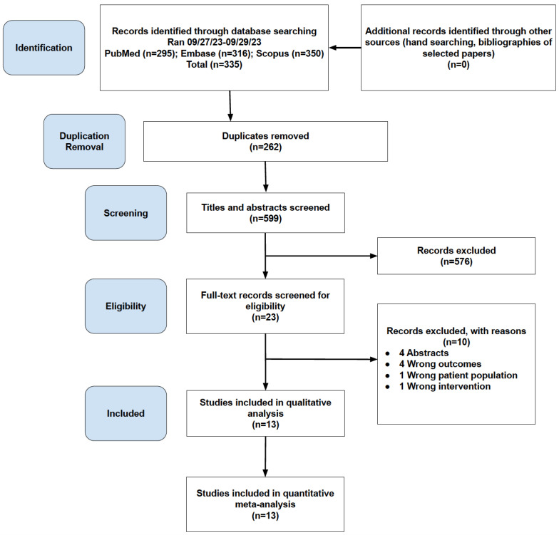
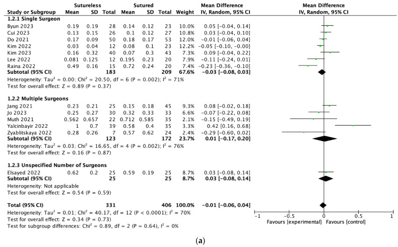
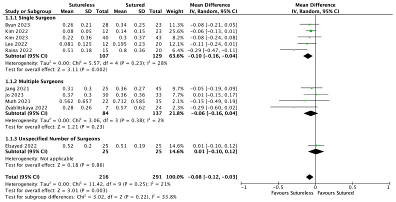
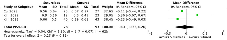
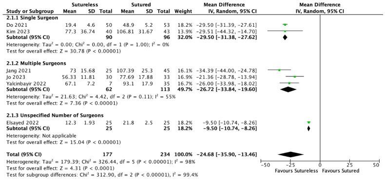
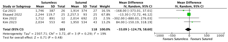
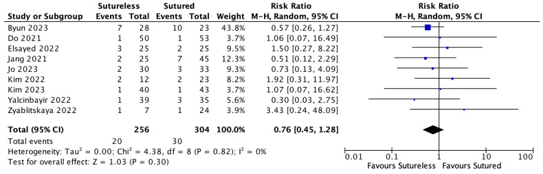
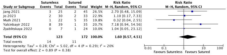
Background: The purpose of the study is to compare the visual outcomes and complications of sutured scleral fixation (SSF), a traditional and conservative surgical approach, and the newer and faster Yamane technique for secondary intraocular lens placement. Methods: A literature search was performed on PubMed, Embase, and Scopus on studies published between 1 July 2017 to 29 September 2023. Outcomes analyzed included the final best corrected visual acuity (BCVA) between 3 and 12 months to assess the effectiveness of the procedure, post-operative month (POM) 1 BCVA to assess the speed of visual recovery, endothelial cell count (ECC), absolute refractive error, surgical duration, and complication rates. Additional subgroup analyses were performed based on surgeon experience with the technique. Single-surgeon studies had an average of 26 procedures performed, whereas multiple-surgeon studies averaged only 9 procedures performed; these were then used to delineate surgeon experience. A sample-size weighted mean difference (MD) meta-analysis was performed across all variables using RevMan 5.4.1; p < 0.05 was considered statistically significant. Results: Thirteen studies with 737 eyes were included: 406 eyes were included in the SSF group, and 331 eyes were included in the Yamane group. There was no significant difference in the final BCVA between groups in both the single-surgeon versus multiple-surgeon studies (MD = -0.01, 95% CI: [-0.06, 0.04], p = 0.73). In the single-surgeon studies, the BCVA at POM1 was significantly improved in the Yamane group compared to SSF (MD = -0.10, 95% CI: [-0.16, -0.04], p = 0.002). In the multiple-surgeon studies, there was no significant difference in BCVA at POM1 (MD = -0.06, 95% CI: [-0.16, 0.04], p = 0.23). The Yamane group had a shorter surgical duration than SSF in both single-surgeon and multiple-surgeon studies (MD = -24.68, 95% CI: [-35.90, -13.46], p < 0.0001). The ECC, refractive error, and complication rates did not significantly differ amongst all groups. Conclusions: The Yamane technique demonstrated similar long-term visual outcomes and complication rates to the traditional SSF. Visual recovery was significantly faster in the Yamane group in the single-surgeon studies. The operative times were shorter across all Yamane groups. Based on these findings, it is advisable to consider the Yamane technique as a viable, and perhaps preferable, option for patients requiring secondary IOL placement, alongside traditional SSF methods.
Keywords: Yamane; flanged haptic; scleral fixation; secondary IOL; sutureless fixation.
Conflict of interest statement
The authors declare that there are no conflicts of interest.
Figures
Similar articles
-
Comparison of three different intraocular lens implantation techniques in the absence of capsular support: sutured scleral, haptic flanged intrascleral, and four flanged intrascleral fixations.Int Ophthalmol. 2023 Dec;43(12):5045-5053. doi: 10.1007/s10792-023-02907-8. Epub 2023 Oct 18. Int Ophthalmol. 2023. PMID: 37851141
-
Comparison of two techniques in posterior lens dislocations: Scleral suture fixation vs. modified Yamane intrascleral lens fixation.J Fr Ophtalmol. 2022 Jan;45(1):13-19. doi: 10.1016/j.jfo.2021.09.009. Epub 2021 Dec 20. J Fr Ophtalmol. 2022. PMID: 34949500
-
Safety and Efficacy of Current Sclera Fixation Methods for Intraocular Lenses and Literature Overview.Klin Monbl Augenheilkd. 2021 Aug;238(8):868-874. doi: 10.1055/a-1333-3199. Epub 2021 Apr 14. Klin Monbl Augenheilkd. 2021. PMID: 33853190 Review. English, German.
-
[Efficacy of sutureless intrascleral intraocular lens fixation with the modified Yamane technique].Zhonghua Yan Ke Za Zhi. 2024 Jun 11;60(6):503-510. doi: 10.3760/cma.j.cn112142-20240103-00008. Zhonghua Yan Ke Za Zhi. 2024. PMID: 38825949 Chinese.
-
Trans-Scleral Plugs Fixated FIL SSF IOL: A Review of the Literature and Comparison with Other Secondary IOL Implants.J Clin Med. 2023 Mar 2;12(5):1994. doi: 10.3390/jcm12051994. J Clin Med. 2023. PMID: 36902780 Free PMC article. Review.
Cited by
-
Comparative Functional and Morphological Data of Different IOL Dislocation Treatment Methods.J Clin Med. 2025 Feb 21;14(5):1462. doi: 10.3390/jcm14051462. J Clin Med. 2025. PMID: 40094913 Free PMC article.
-
Scleral-fixated IOLs - A comprehensive review of current practices and emerging trends.Indian J Ophthalmol. 2025 Jul 1;73(7):933-945. doi: 10.4103/IJO.IJO_2812_24. Epub 2025 Jun 30. Indian J Ophthalmol. 2025. PMID: 40586185 Free PMC article. Review.
-
Timing of the modified yamane technique in complicated cataract surgery: same session or second session?Int Ophthalmol. 2025 May 19;45(1):205. doi: 10.1007/s10792-025-03571-w. Int Ophthalmol. 2025. PMID: 40387987
References
-
- Czajka M.P., Frajdenberg A., Stopa M., Pabin T., Johansson B., Jakobsson G. Sutureless intrascleral fixation using different three-piece posterior chamber intraocular lenses: A literature review of surgical techniques in cases of insufficient capsular support and a retrospective multicentre study. Acta Ophthalmol. 2020;98:224–236. doi: 10.1111/aos.14307. - DOI - PubMed
Publication types
LinkOut - more resources
Full Text Sources
