. 2024 Jun 3;13(11):3293.
doi: 10.3390/jcm13113293.

Croco Eye Technique: Mucous Retention Cyst Excision with Immediate Open Sinus Lift-A Retrospective Cohort Study

Affiliations

Croco Eye Technique: Mucous Retention Cyst Excision with Immediate Open Sinus Lift-A Retrospective Cohort Study

Radosław Jadach et al. J Clin Med. .

Abstract

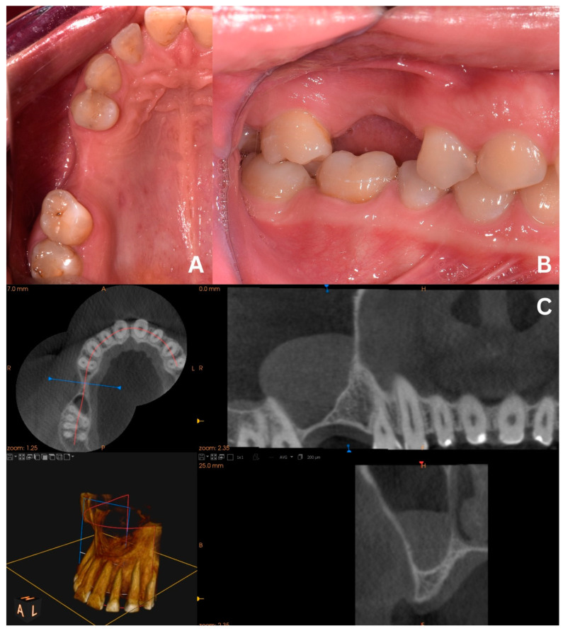
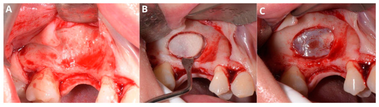
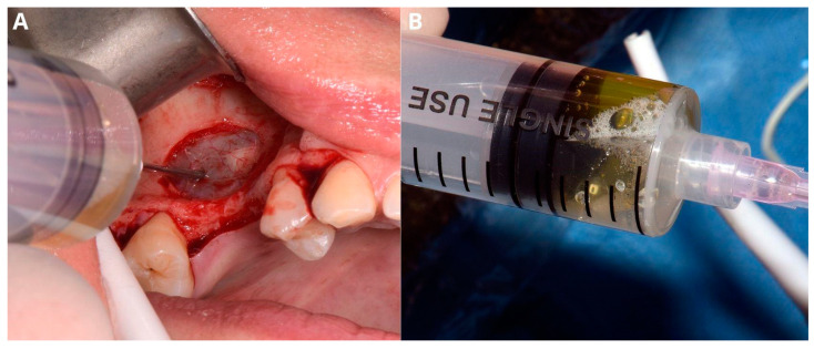
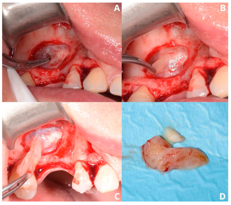
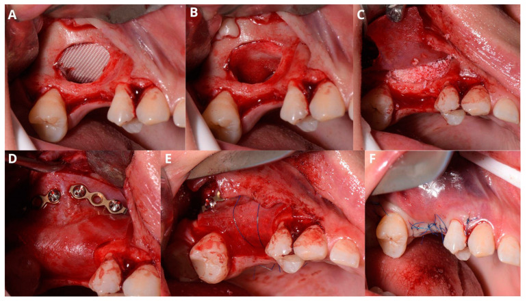
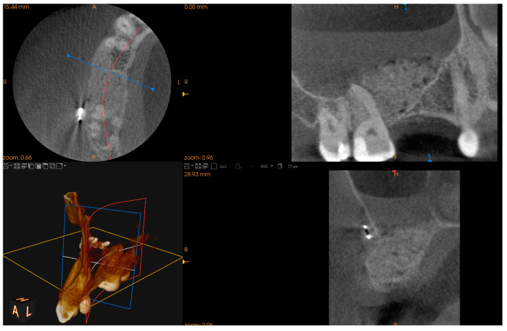
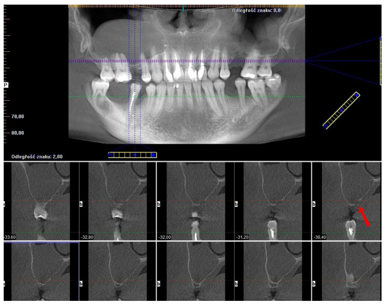
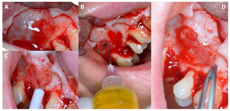
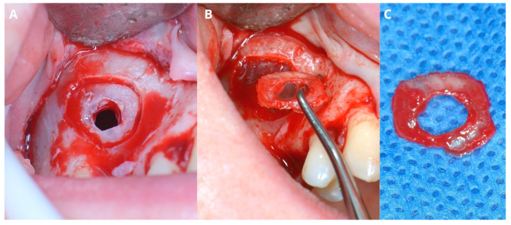
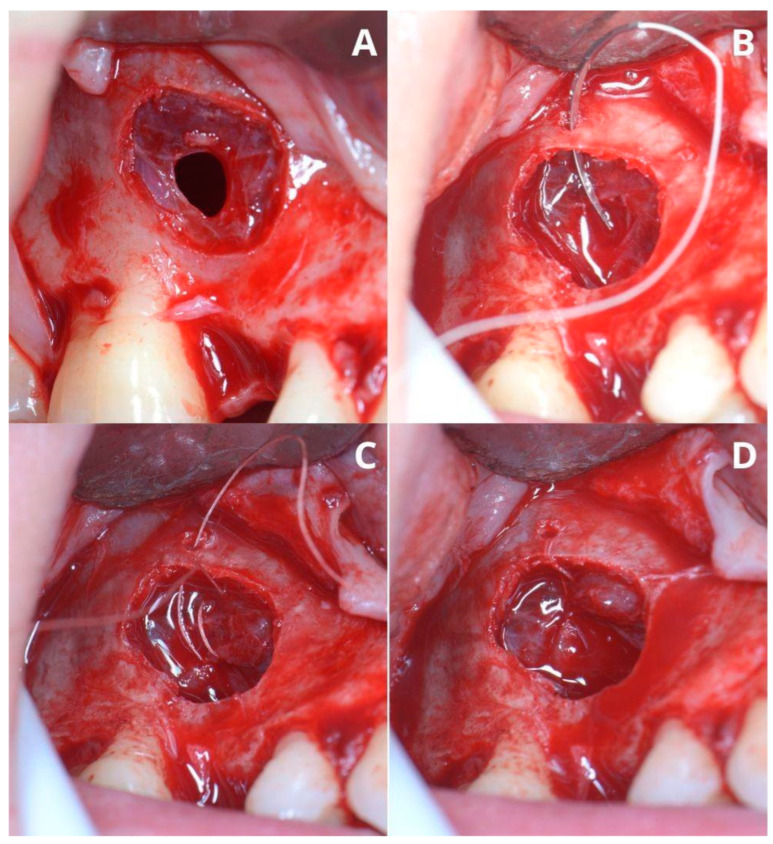
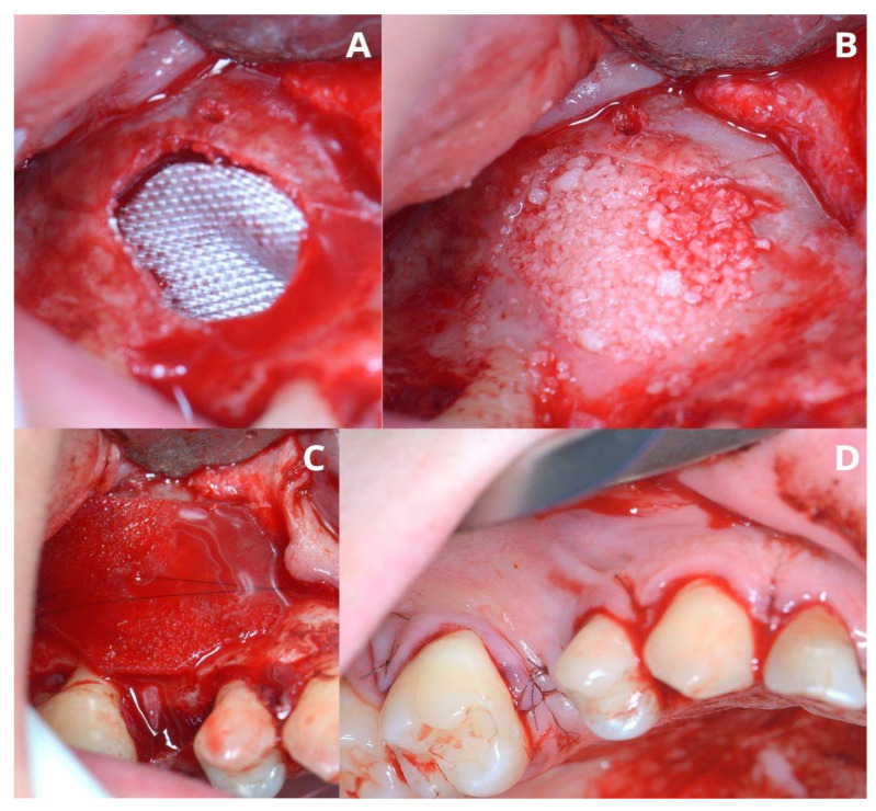
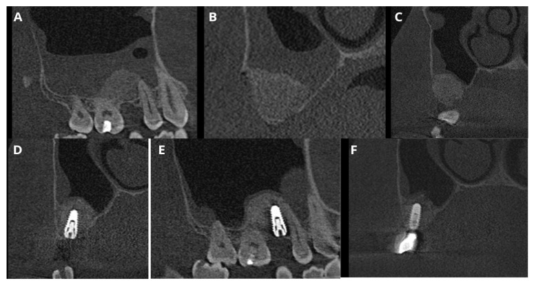
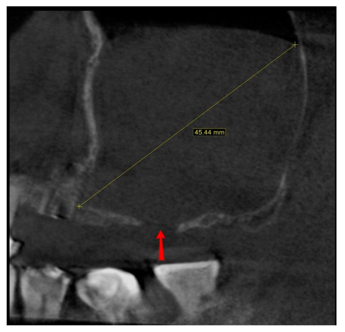
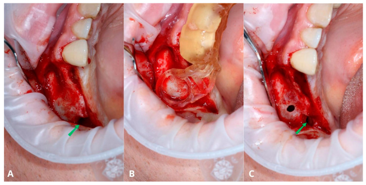
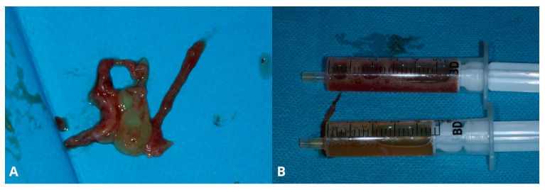
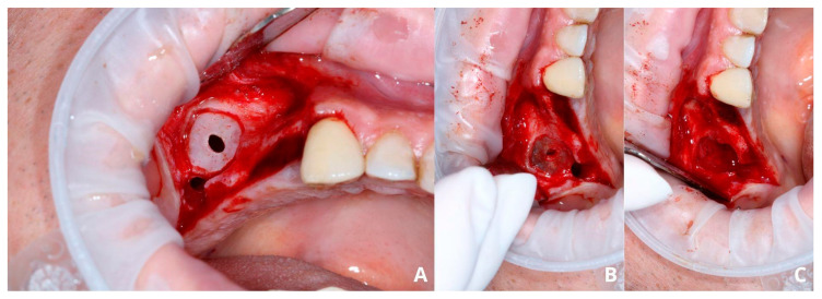
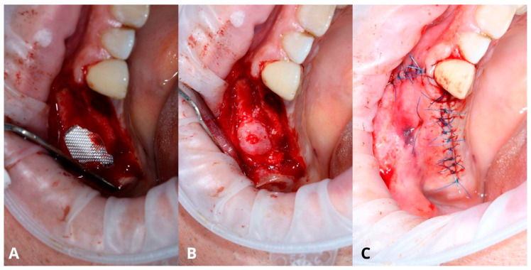
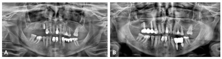
Objectives: A mucous retention cyst is a common, asymptomatic lesion that may cause complications during or after the sinus lift procedure. The goal of this study is to assess the effectiveness of the Croco Eye Technique (CET), which allows simultaneous excision of the cyst and sinus floor elevation. Methods: The technique was thoroughly described in two versions, and the group of 33 patients was analyzed. Patients who qualified for this procedure had insufficient alveolar ridge height, and their CBCT showed radiological images typical for retention cysts. Analyzed parameters included the version of CET, demographic data, anatomical parameters, intraoperative complications, recurrence of the cyst, success rate of the sinus lift and implants, and the follow-up period. Results: Out of the 33 cases, 9 were of the primary version (27.27%) and 24 of the final version (72.73%). The average height of a retention cyst was 24.05 mm, with the average alveolar ridge height of 1.86 mm. In three cases (9.09%), implants were placed immediately. The prevalence of uncontrolled Schneiderian membrane perforation was reduced from 55.56% to 4.17% between the primary and final versions. The cyst's recurrence rate was 3.13%. The implant survival rate was 100%. The mean follow-up period was 48.625 months (max 110 months). Conclusions: The Croco Eye Technique, despite the perforation of the Schneiderian membrane, enables successful sinus lift and implantation with a success rate of 100%. Excision of the retention cyst, which is the cause of perforation, allows for limiting the risk of the cyst's recurrence.

Keywords: biopsy; croco eye technique; excision; mucous retention cyst; retrospective cohort study; sinus floor elevation; sinus lift.


Conflict of interest statement

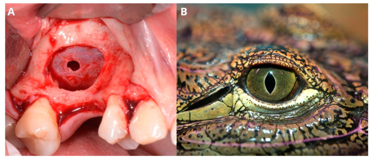
The authors declare no conflicts of interest. The authors report no proprietary or commercial interest in any product mentioned or concept discussed in this article. The photography of a crocodile eye (Figure 5B) was purchased via Canva Pro (Canva Pty Ltd., Perth, Australia).

Figures

Figure 1
Figure 1
Clinical situation before the surgery. Occlusal view (A), lateral view (B), and CBCT scans with a dome-shaped radiopaque lesion in the sinus and insufficient bone height that requires sinus lift before placing implants (C).
Figure 2
Figure 2
Creating access through the lateral window. Elevation of the full-thickness flap (A), osteotomy with piezotome, and removal of the “bony lid” (B), undamaged Schneiderian membrane with visible vasculature (C).
Figure 3
Figure 3
Aspiration of the fluid from the retention cyst. The needle punctures the cyst in the center of the bone window (A), which facilitates management of the future perforation; yellowish color of the retention cyst’s content (B).
Figure 4
Figure 4
Removal of the cysts’s lining. Two layers of the Schneiderian membrane are visible: periosteum and mucosa (A). Gentle removal of the lining (B,C) and its remains (D).
Figure 5
Figure 5
Perforated Schneiderian membrane (A) resembles the eye of a crocodile (B) (photography purchased via Canva Pro (Canva Pty Ltd., Perth, Australia)).
Figure 6
Figure 6
Completing the sinus lift. The first layer that was put under the Schneiderian membrane was oxycellulose (hemostatic material BloodSTOP™ iX (LifeScience Plus, Mountain View, CA, USA)) (A). Then, a thick, shape-retaining collagen membrane (B) with a xenograft was placed below it (C). The second membrane (to rebuild the bone horizontally) was fixed with two osseofixation plates (D) and sutured to the palate soft tissues (nylon 5-0) (E) to obtain tension-free contact with the flap’s edges. Final suturing was performed with nylon 5-0 (F).
Figure 7
Figure 7
Screenshot from post-op CBCT scans with visible xenograft and a flat level of liquid in the maxillary sinus lumen due to present perforation.
Figure 8
Figure 8
Croco Eye Technique—final version. Screenshot of the pre-op CBCT scans with a visible radiopaque lesion in the right maxillary sinus, insufficient alveolar ridge height, and noncontinuous bone of the sinus floor due to past oroantral communication (OAC) (red arrow).
Figure 9
Figure 9
Preparing for the first osteotomy. Elevation of a full-thickness flap (A). Creating a smaller window and aspirating the retention cyst’s yellow content (B). Removal of the cyst’s lining, firstly with narrow suction (C), then gently with tweezers (D). The perforation will not enlarge more than the diameter of osteotomy, as the periosteum is still attached to the bone, therefore remains safe.
Figure 10
Figure 10
Creating the second, larger window around the perforation (A). Detachment of the bony ring from the Schneiderian membrane (B) and the ring itself (C).
Figure 11
Figure 11
Managing the Schneiderian membrane perforation. The perforation is located in the center of the bigger window (A). The needle of the resorbable PGA (polyglycolic acid) 5-0 suture gets through a tiny hole (which was previously drilled) in the upper edge of the window (B). A double loop closes the perforation (C), and the knot is made around the tiny bone hole (D).
Figure 12
Figure 12
Completing the sinus lift. Oxycellulose was put directly under the Schneiderian membrane (A), then xenografted with I-PRF (B) and closed with collagen membrane (C). The horizontal suture (5-0 nylon) is meant to hold the collagen membrane in place and prevent the xenograft from migrating buccally to the soft tissues. The wound was closed with 5-0 nylon sutures (D).
Figure 13
Figure 13
Screenshots of the post-op CBCT scans. Immediately after Croco Eye (A,B), 6 months post-op: before (C) and after implant placement (D,E). The latest follow-up was 5 years 2 months post-op with a loaded implant (F). Throughout the follow-up period, no recurrence of the cyst was detected.
Figure 14
Figure 14
Screenshot from the pre-op CBCT scan showing maximal retention cyst’s diameter (45.44 mm; yellow line). Also, the past oroantral communication window is visible (red arrow).
Figure 15
Figure 15
A 3D-printed surgical guide was created to simplify the osteotomy and to determine an optimal location for placing a xenograft during sinus lift.
Figure 16
Figure 16
Full-thickness flap elevation with an oroantral communication (OAC) (green arrow) visible from the very beginning of the surgery (A). The rounded part of the surgical guide indicates the most optimal location of the osteotomy from the augmentation point of view (B). Smaller osteotomy after removal of the cyst’s lining (C). The OAC is also visible here (green arrow).
Figure 17
Figure 17
The extracted cyst’s lining (A) and yellowish content were drawn with syringes (B).
Figure 18
Figure 18
The second osteotomy was created around the first one (A). Situation after the removal of the bony lid (B) and after the elevation of the Schneiderian membrane with visible resorbable suture (C).
Figure 19
Figure 19
Oxycellulose was placed just under the Schneiderian membrane (A) and then xenografted with I-PRF, collagen, and A-PRF membranes. The bony lid was put back in place (B) and later stabilized with a mattress suture. Final closure with 5-0 nylon sutures (C).
Figure 20
Figure 20
Comparison of orthopantomogram (OPG) immediately after the procedure (A) and 4 years after delivering the final reconstruction on implants (B).

Similar articles

Cited by

References

    1. Hung K., Hui L., Yeung A.W.K., Wu Y., Hsung R.T.-C., Bornstein M.M. Volumetric analysis of mucous retention cysts in the maxillary sinus: A retrospective study using cone-beam computed tomography. Imaging Sci. Dent. 2021;51:117–127. doi: 10.5624/isd.20200267. - DOI - PMC - PubMed
    1. Anitua E., Alkhraisat M., Torre A., Eguia A. Are mucous retention cysts and pseudocysts in the maxillary sinus a risk factor for dental implants? A systematic review. Med. Oral Patol. Oral Cir. Bucal. 2021;26:e276–e283. doi: 10.4317/medoral.24155. - DOI - PMC - PubMed
    1. Chan H.-L., Wang H.-L. Sinus pathology and anatomy in relation to complications in lateral window sinus augmentation. Implant Dent. 2011;20:406–412. doi: 10.1097/ID.0b013e3182341f79. - DOI - PubMed
    1. Berberi A., Aad G., Kebbe S., El Hachem R., Nader N. Treatment of Mucous Retention Cyst in Association with Sinus Lift and Implant Placement: A Case Report with 1-Year Follow-Up. Case Rep. Dent. 2023;2023:6968487. doi: 10.1155/2023/6968487. - DOI - PMC - PubMed
    1. Aghaee F., Moudi E., Vahdani N., Bijani A., Haghanifar S. Evaluation of Anatomical Variations of the Maxillary Sinus in Patients with and Without Mucous Retention Cyst. Ear Nose Throat J. 2023 doi: 10.1177/01455613231206284. - DOI - PubMed

LinkOut - more resources