. 2024 Jun 6;13(11):3348.
doi: 10.3390/jcm13113348.

All-on-4 Hybrid with Extra-Long Transnasal Implants: Descriptions of the Technique and Short-Term Outcomes in Three Cases

Affiliations

All-on-4 Hybrid with Extra-Long Transnasal Implants: Descriptions of the Technique and Short-Term Outcomes in Three Cases

Mariana Nunes et al. J Clin Med. .

Abstract

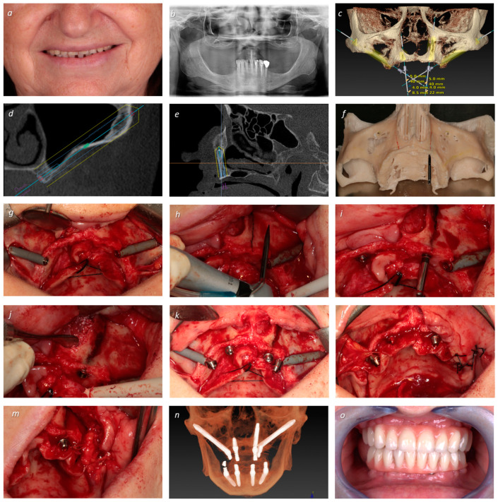
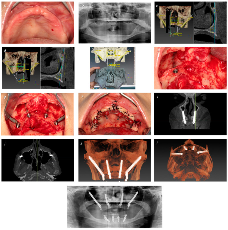
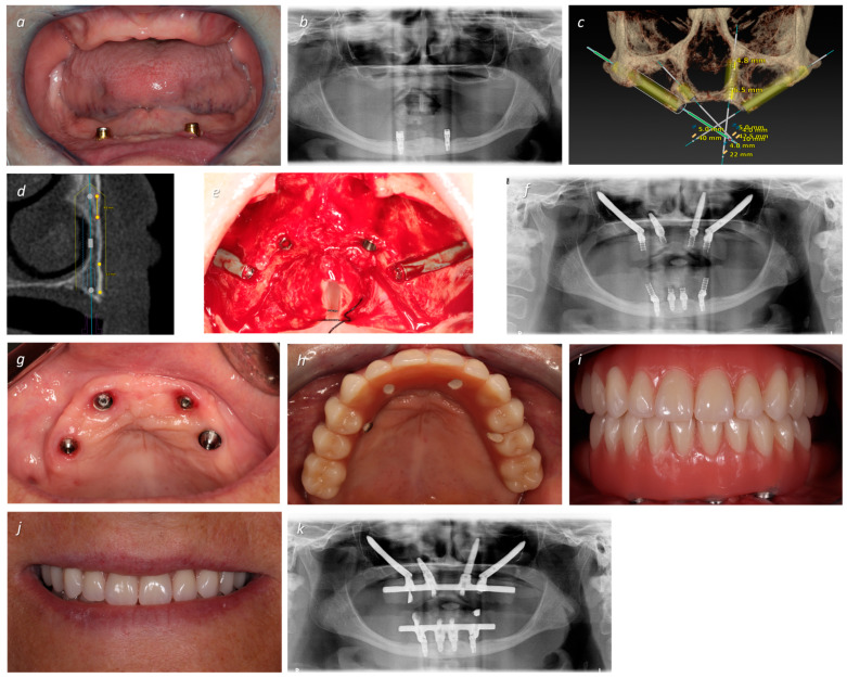
Background/Objectives: There is a need for alternative approaches to full-arch rehabilitation of atrophic maxillae. The aim of this short case series was to describe the technique and assess the short term-outcomes of atrophic maxillae rehabilitation using transnasal implants in conjunction with zygomatic implants. Methods: Three female patients (average age: 62 years) presenting comorbidities and atrophic maxillae preventing the insertion of standard maxillary anchored implants received a full-arch fixed prosthesis supported by transnasal implants together with zygomatic implants, using the ad modum all-on-4 concept. Patients were followed during the functional osseointegration period. Primary outcome measures were prosthetic and implant survival based on function. Secondary outcome measures were complication parameters (biological and mechanical), plaque and bleeding levels, and probing pocket depths > 4 mm. Results: No implant failures were registered, and all prostheses remained in function. The only complication was a fracture of a provisional crown that was resolved. Plaque and bleeding scores were mild during the follow-up period. Conclusions: The present manuscript describes the use of extra-long transnasal implants in combination with zygomatic implants in immediate function for full-arch fixed prosthetic rehabilitation of atrophic maxillae, with the objective of promoting more research into this relatively recent technique. More studies are needed to validate the technique.

Keywords: all-on-4; dental implants; transnasal; zygomatic implants.


Conflict of interest statement

The authors declare no conflicts of interest.

Figures

Figure 1
Figure 1
A 66-year-old female patient with antidepressant and anxiolytic medication complained of total missing teeth in upper jaw using a removable denture for 15 years. (a) Clinical examination revealed large resorption and loss of VOD; (b) orthopantomography revealing extensive bone loss on the maxilla with high pneumatization in both sites; (c) virtual implant planning, placing zygomatic implants (positions #15 and #25) combined with one conventional (position #12) and one transnasal implant (position #22); (d) virtual zygomatic implant planning; (e) virtual transnasal implant planning (NASA 3 classification) [24] (note the nasolacrimal canal); (f) stereolithographic model planning; (g) intra-oral peri-operative view with insertion of two NobelZygoma 0° with 40 mm of length (Nobel Biocare AB) in positions #15 and #25; (h) drilling pathway for the transnasal implant preparation; (i) insertion of the transnasal implant in position #22 (Helix GM® Long 3.75 × 22.5 mm, Neodent, Curitiba, Brazil), where less than 4 mm of bone was available; (j) performing a bone graft (Creos, Nobel Biocare AB) along the internal extra-osseous portion of the transnasal implant; (k) intra-oral peri-operative view after connection of multi-unit abutments (Nobel Biocare AB): 30° and 5 mm of height (zygomatic implant in position #15), 45° and 6 mm of height (zygomatic implant in position #25), 17° and 3 mm of height (standard implant in position #12), 30° and 2.5 mm of height (transnasal implant in position #22); Bichat ball used as graft over the extra-osseous portion of the zygomatic implants: (l) implant in position #15; (m) implant in position #25; (n) post-operative CBCT displaying the bimaxillary rehabilitations performed in the immediate function; (o) frontal post-operative view of the rehabilitations with connection of the provisional prosthesis on the same day of surgery, achieving immediate function.
Figure 2
Figure 2
A 56-year-old female patient with musculoskeletal disease undergoing treatment with physiotherapy and analgesic medication, complaining of painful removable prosthesis and absolute loss of retention. (a) Intra-oral pre-operative occlusal photograph of the maxilla exhibiting significant resorption; (b) orthopantomography revealing significant resorption and high sinus pneumatization disabling the insertion of short implants in the anterior maxilla; (c) transnasal right implant planning in position #12 (NASA 3 classification) [24] (note also the planned position of the right zygomatic implant in position #15); (d) transnasal left implant planning in position #22 (NASA 3 classification) [24] (note also the planned position of the left zygomatic implant in position #25); (e) surgical simulation on the stereolithographic model (prototype of the middle third of the face with extended maxilla); (f) intra-oral peri-operative view of the right transnasal implant placed, and the left transnasal implant pathway marked with a pencil; (g) intra-oral peri-operative view with insertion of two NobelZygoma 0° (Nobel Biocare AB) in positions #15 (40 mm of length) and #25 (40 mm of length), and two transnasal implants in positions #12 and #22 (Helix GM® Long 3.75 × 22.5 mm, Neodent, Curitiba, Brazil) with all multi-unit abutments connected; (h) intra-oral peri-operative photograph after suturing and before impressions, with the impression copings attached to the abutments; (i) post-operative CBCT slice exhibiting the final frontal transnasal implant’s position; (j) post-operative CBCT slice exhibiting the axial view of the final transnasal apex implant’s position; (k) post-operative CBCT frontal view of the final implant rehabilitation; (l) post-operative CBCT axial view of the final implant rehabilitation; (m) post-operative CBCT of the final bimaxillary rehabilitation achieving immediate function.
Figure 3
Figure 3
A 64-year-old female patient with hypertension and a history of psychological complications, with lack of retention of both removable prostheses. (a) Frontal pre-operative intra-oral photograph (note the large resorption and loss of VOD); (b) pre-operative orthopantomography revealing severe bone loss and inadequate bone in the maxillary sinus and anterior region; (c) virtual implant planning, placing zygomatic implants (positions #15 and #25) combined with one conventional mesially-tilted implant (position #12), aiming for increased bone contact surface, and one transnasal implant (position #22); (d) transnasal implant planning with the DTX software (V3.6.4.2; NASA 3 classification) [24], where CBCT slices reveal that this implant is anchored in 6.5 mm of the crest and 4.8 mm of the nasal concha; (e) intra-oral peri-operative view with insertion of two NobelZygoma 0° with 40 mm of length (Nobel Biocare AB) in positions #15 and #25, one NobelSpeedy Groovy RP 10 mm (Nobel Biocare AB) in position #12, and one Helix GM® Long 3.75 × 22.5 mm (Neodent, Curitiba, Brazil) in position #22; (f) post-operative orthopantomography displaying the bimaxillary rehabilitations performed in immediate function; (g) occlusal post-operative intra-oral photograph displaying the healing at 4 months of follow-up; (h) occlusal intra-oral photograph displaying the definitive prosthesis (a titanium infrastructure with acrylic resin artificial gingiva and crowns); (i) frontal intra-oral photograph displaying the definitive prosthesis; (j) patient smiling with the definitive prosthesis; (k) final orthopantomography with the bimaxillary rehabilitations.

Similar articles

References

    1. World Health Organization . World Health Organization; 2023. [(accessed on 24 March 2024)]. Oral Health. Available online: https://www.who.int/health-topics/oral-health#tab=tab_1.
    1. Atwood D.A. Reduction of residual ridges: A major oral disease entity. J. Prosthet. Dent. 1971;26:266–279. doi: 10.1016/0022-3913(71)90069-2. - DOI - PubMed
    1. Maló P., de Araújo Nobre M., Lopes A., Ferro A., Nunes M. The All-on-4 concept for full-arch rehabilitation of the edentulous maxillae: A longitudinal study with 5–13 years of follow-up. Clin. Implant. Dent. Relat. Res. 2019;21:538–549. doi: 10.1111/cid.12771. - DOI - PubMed
    1. Maló P., de Araújo Nobre M., Lopes A., Ferro A., Botto J. The All-on-4 treatment concept for the rehabilitation of the completely edentulous mandible: A longitudinal study with 10 to 18 years of follow-up. Clin. Implant. Dent. Relat. Res. 2019;21:565–577. doi: 10.1111/cid.12769. - DOI - PubMed
    1. Ferro A.S., de Araújo Nobre M.A., Simões R. Ten-year follow-up of full-arch rehabilitations supported by implants in immediate function with nasal and full-length palatine bicortical anchorage on the anterior maxilla. J. Oral Sci. 2022;64:129–134. doi: 10.2334/JOSNUSD.21-0378. - DOI - PubMed

LinkOut - more resources