

Quantitative Bone SPECT/CT of Central Cartilaginous Bone Tumors: Relationship between SUVmax and Radiodensity in Hounsfield Unit
- PMID: 38893090
- PMCID: PMC11171356
- DOI: 10.3390/cancers16111968
Quantitative Bone SPECT/CT of Central Cartilaginous Bone Tumors: Relationship between SUVmax and Radiodensity in Hounsfield Unit
Abstract
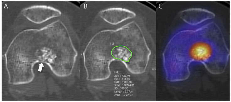
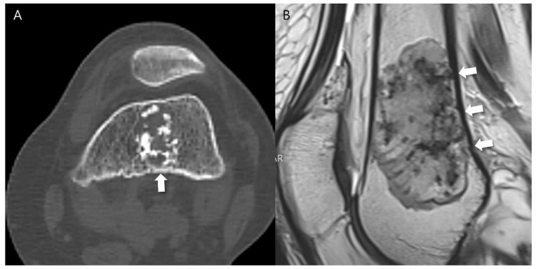
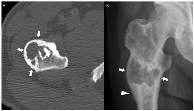
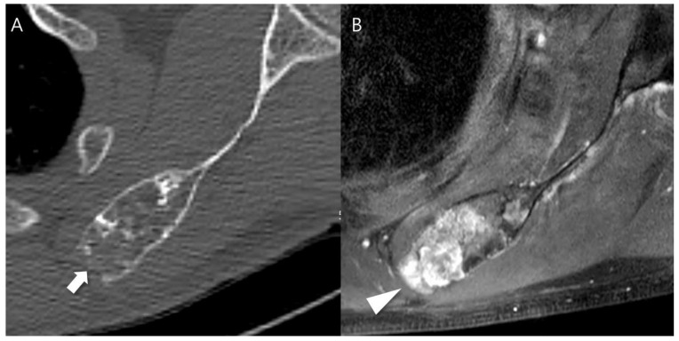
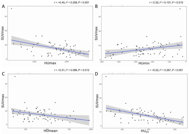
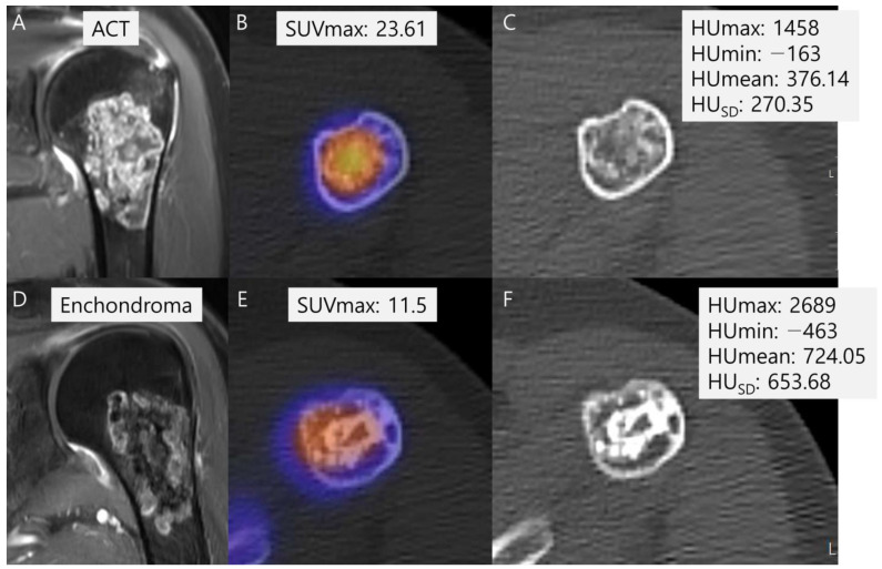
(1) Background: it is challenging to determine the accurate grades of cartilaginous bone tumors. Using bone single photon emission computed tomography (SPECT)/computed tomography (CT), maximum standardized uptake value (SUVmax) was found to be significantly associated with different grades of cartilaginous bone tumor. The inquiry focused on the effect of the tumor matrix on SUVmax. (2) Methods: a total of 65 patients from 2017 to 2022 with central cartilaginous bone tumors, including enchondromas and low-to-intermediate grade chondrosarcomas, who had undergone bone SPECT/CT were retrospectively enrolled. The SUVmax was recorded and any aggressive CT findings of cartilaginous bone tumor and Hounsfield units (HU) of the chondroid matrix as mean, minimum, maximum, and standard deviation (SD) were reviewed on CT scans. Pearson's correlation analysis was performed to determine the relationship between CT features and SUVmax. Subgroup analysis was also performed between the benign group (enchondroma) and the malignant group (grade 1 and 2 chondrosarcoma) for comparison of HU values and SUVmax. (3) Results: a significant negative correlation between SUVmax and HU measurements, including HUmax, HUmean, and HUSD, was found. The subgroup analysis showed significantly higher SUVmax in the malignant group, with more frequent CT aggressive features, and significantly lower HUSD in the malignant group than in the benign group. (4) Conclusions: it was observed that higher SUVmax and lower HUSD were associated with a higher probability of having a low-to-intermediate chondrosarcoma with aggressive features and a less calcified tumor matrix.
Keywords: SUVmax; bone SPECT/CT; cartilaginous bone tumor; chondroid matrix mineralization; correlation; hounsfield units.
Conflict of interest statement
The authors declare no conflicts of interest.
Figures
Similar articles
-
Correlation between the maximum standard uptake value and mean Hounsfield unit on single-photon emission computed tomography-computed tomography to discriminate benign and metastatic lesions among patients with breast cancer.Asian Spine J. 2024 Jun;18(3):398-406. doi: 10.31616/asj.2022.0451. Epub 2024 Jun 25. Asian Spine J. 2024. PMID: 38917860 Free PMC article.
-
Quantitative SPECT/CT for differentiating between enchondroma and grade I chondrosarcoma.Sci Rep. 2020 Jun 29;10(1):10587. doi: 10.1038/s41598-020-67506-4. Sci Rep. 2020. PMID: 32601314 Free PMC article.
-
Standardized uptake values of 99mTc-MDP in normal vertebrae assessed using quantitative SPECT/CT for differentiation diagnosis of benign and malignant bone lesions.BMC Med Imaging. 2021 Feb 27;21(1):39. doi: 10.1186/s12880-021-00569-5. BMC Med Imaging. 2021. PMID: 33639883 Free PMC article.
-
The utility of 18F-FDG PET and PET/CT in the diagnosis and staging of chondrosarcoma: a meta-analysis.J Orthop Surg Res. 2020 Jun 22;15(1):229. doi: 10.1186/s13018-020-01748-w. J Orthop Surg Res. 2020. PMID: 32571371 Free PMC article.
-
Chondroid Tumors as Incidental Findings and Differential Diagnosis between Enchondromas and Low-grade Chondrosarcomas.Semin Musculoskelet Radiol. 2019 Feb;23(1):3-18. doi: 10.1055/s-0038-1675550. Epub 2019 Jan 30. Semin Musculoskelet Radiol. 2019. PMID: 30699449 Review.
Cited by
-
Whole-body imaging for distant staging of bone chondrosarcoma: a systematic review.Radiol Med. 2025 Jul 23. doi: 10.1007/s11547-025-02054-3. Online ahead of print. Radiol Med. 2025. PMID: 40699280 Review.
References
-
- WHO Classification of Tumours Editorial Board . WHO Classification of Tumours: Soft Tissue and Bone Tumours. International Agency for Research on Cancer; Lyon, France: 2020.
Grants and funding
LinkOut - more resources
Full Text Sources
