. 2024 May 25;16(11):2010.
doi: 10.3390/cancers16112010.

Attenuation of the BOLD fMRI Signal and Changes in Functional Connectivity Affecting the Whole Brain in Presence of Brain Metastasis

Affiliations

Attenuation of the BOLD fMRI Signal and Changes in Functional Connectivity Affecting the Whole Brain in Presence of Brain Metastasis

Pia Angstwurm et al. Cancers (Basel). .

Abstract

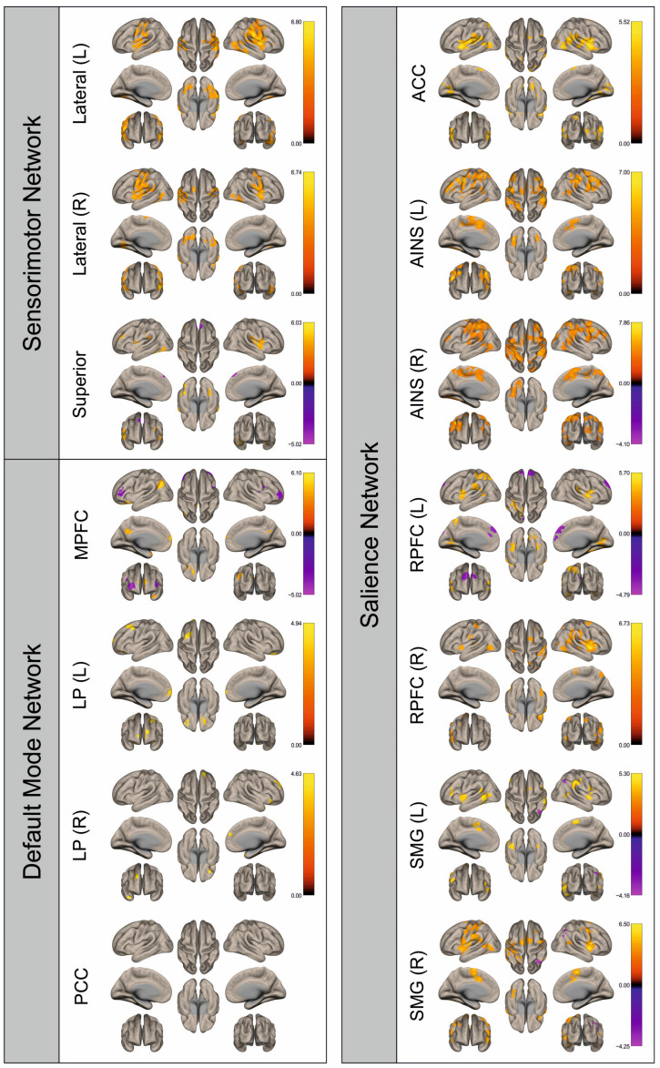
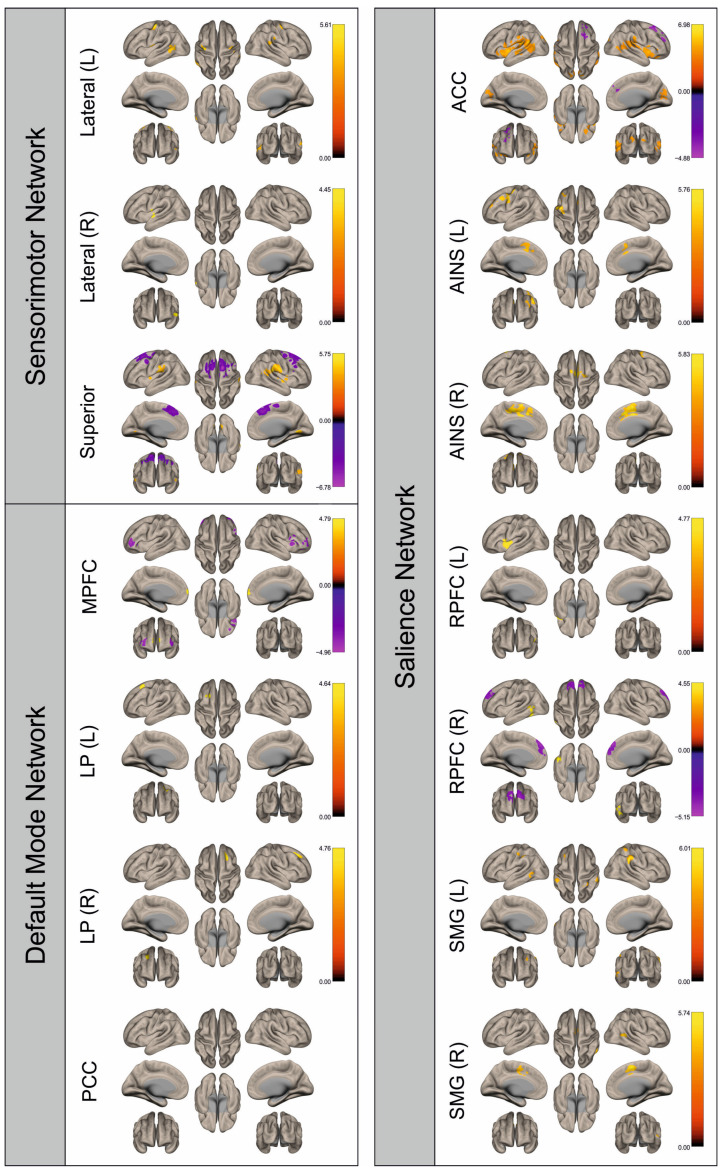
To date, there are almost no investigations addressing functional connectivity (FC) in patients with brain metastases (BM). In this retrospective study, we investigate the influence of BM on hemodynamic brain signals derived from functional magnetic resonance imaging (fMRI) and FC. Motor-fMRI data of 29 patients with BM and 29 matched healthy controls were analyzed to assess percent signal changes (PSC) in the ROIs motor cortex, premotor cortex, and supplementary motor cortex and FC in the sensorimotor, default mode, and salience networks using Statistical Parametric Mapping (SPM12) and marsbar and CONN toolboxes. In the PSC analysis, an attenuation of the BOLD signal in the metastases-affected hemisphere compared to the contralateral hemisphere was significant only in the supplementary motor cortex during hand movement. In the FC analysis, we found alterations in patients' FC compared to controls in all examined networks, also in the hemisphere contralateral to the metastasis. This indicates a qualitative attenuation of the BOLD signal in the affected hemisphere and also that FC is altered by the presence of BM, similarly to what is known for primary brain tumors. This transformation is not only visible in the infiltrated hemisphere, but also in the contralateral one, suggesting an influence of BM beyond local damage.

Keywords: blood oxygenation level-dependent imaging; brain metastasis; functional connectivity; functional magnetic resonance imaging; percent signal change.


Conflict of interest statement

The authors declare no conflicts of interest.

Figures

Figure 1
Figure 1
Percent signal changes of the examined Regions of Interest within the patient group for affected vs. the unaffected hemispheres. Statistically significant results are marked with * for p < 0.05.
Figure 2
Figure 2
Surface maps showing significant differences in functional connectivity between the patients with a left hemispheric metastasis and their matched controls for the paradigm of the left hand. Areas shown in red to yellow are more strongly connected to the examined seeds in controls, while areas shown in blue to purple are more strongly connected to the seeds in patients. Abbreviations: L: left, R: right, MPFC: medial prefrontal cortex, LP: lateral parietal, PCC: posterior cingulate cortex, ACC: anterior cingulate cortex, AINS: anterior insula, RPFC: rostral prefrontal cortex, SMG: supramarginal gyrus.
Figure 3
Figure 3
Surface maps showing significant differences in functional connectivity between the patients with a left hemispheric metastasis and their matched controls for the paradigm of the right hand. Areas shown in red to yellow are more strongly connected to the examined seeds in controls, while areas shown in blue to purple are more strongly connected to the seeds in patients. Abbreviations: L: left, R: right, MPFC: medial prefrontal cortex, LP: lateral parietal, PCC: posterior cingulate cortex, ACC: anterior cingulate cortex, AINS: anterior insula, RPFC: rostral prefrontal cortex, SMG: supramarginal gyrus.
Figure 4
Figure 4
Surface maps showing significant differences in functional connectivity between the patients with a left hemispheric metastasis and their matched controls for the paradigm of the left foot. Areas shown in red to yellow are more strongly connected to the examined seeds in controls, while areas shown in blue to purple are more strongly connected to the seeds in patients. Abbreviations: L: left, R: right, MPFC: medial prefrontal cortex, LP: lateral parietal, PCC: posterior cingulate cortex, ACC: anterior cingulate cortex, AINS: anterior insula, RPFC: rostral prefrontal cortex, SMG: supramarginal gyrus.
Figure 5
Figure 5
Surface maps showing significant differences in functional connectivity between the patients with a left hemispheric metastasis and their matched controls for the paradigm of the right foot. Areas shown in red to yellow are more strongly connected to the examined seeds in controls, while areas shown in blue to purple are more strongly connected to the seeds in patients. Abbreviations: L: left, R: right, MPFC: medial prefrontal cortex, LP: lateral parietal, PCC: posterior cingulate cortex, ACC: anterior cingulate cortex, AINS: anterior insula, RPFC: rostral prefrontal cortex, SMG: supramarginal gyrus.
Figure 6
Figure 6
Surface maps showing significant differences in functional connectivity between the patients with a right hemispheric metastasis and their matched controls for the paradigm of the left hand. Areas shown in red to yellow are more strongly connected to the examined seeds in controls, while areas shown in blue to purple are more strongly connected to the seeds in patients. Abbreviations: L: left, R: right, MPFC: medial prefrontal cortex, LP: lateral parietal, PCC: posterior cingulate cortex, ACC: anterior cingulate cortex, AINS: anterior insula, RPFC: rostral prefrontal cortex, SMG: supramarginal gyrus.
Figure 7
Figure 7
Surface maps showing significant differences in functional connectivity between the patients with a right hemispheric metastasis and their matched controls for the paradigm of the right hand. Areas shown in red to yellow are more strongly connected to the examined seeds in controls, while areas shown in blue to purple are more strongly connected to the seeds in patients. Abbreviations: L: left, R: right, MPFC: medial prefrontal cortex, LP: lateral parietal, PCC: posterior cingulate cortex, ACC: anterior cingulate cortex, AINS: anterior insula, RPFC: rostral prefrontal cortex, SMG: supramarginal gyrus.
Figure 8
Figure 8
Surface maps showing significant differences in functional connectivity between the patients with a right hemispheric metastasis and their matched controls for the paradigm of the left foot. Areas shown in red to yellow are more strongly connected to the examined seeds in controls, while areas shown in blue to purple are more strongly connected to the seeds in patients. Abbreviations: L: left, R: right, MPFC: medial prefrontal cortex, LP: lateral parietal, PCC: posterior cingulate cortex, ACC: anterior cingulate cortex, AINS: anterior insula, RPFC: rostral prefrontal cortex, SMG: supramarginal gyrus.
Figure 9
Figure 9
Surface maps showing significant differences in functional connectivity between the patients with a right hemispheric metastasis and their matched controls for the paradigm of the right foot. Areas shown in red to yellow are more strongly connected to the examined seeds in controls, while areas shown in blue to purple are more strongly connected to the seeds in patients. Abbreviations: L: left, R: right, MPFC: medial prefrontal cortex, LP: lateral parietal, PCC: posterior cingulate cortex, ACC: anterior cingulate cortex, AINS: anterior insula, RPFC: rostral prefrontal cortex, SMG: supramarginal gyrus.

Similar articles

Cited by

References

    1. Gállego Pérez-Larraya J., Hildebrand J. Brain metastases. Handb. Clin. Neurol. 2014;121:1143–1157. doi: 10.1016/B978-0-7020-4088-7.00077-8. - DOI - PubMed
    1. Ostrom Q.T., Wright C.H., Barnholtz-Sloan J.S. Brain metastases: Epidemiology. Handb. Clin. Neurol. 2018;149:27–42. doi: 10.1016/B978-0-12-811161-1.00002-5. - DOI - PubMed
    1. Cagney D.N., Martin A.M., Catalano P.J., Redig A.J., Lin N.U., Lee E.Q., Wen P.Y., Dunn I.F., Bi W.L., Weiss S.E., et al. Incidence and prognosis of patients with brain metastases at diagnosis of systemic malignancy: A population-based study. Neuro Oncol. 2017;19:1511–1521. doi: 10.1093/neuonc/nox077. - DOI - PMC - PubMed
    1. Moravan M.J., Fecci P.E., Anders C.K., Clarke J.M., Salama A.K.S., Adamson J.D., Floyd S.R., Torok J.A., Salama J.K., Sampson J.H., et al. Current multidisciplinary management of brain metastases. Cancer. 2020;126:1390–1406. doi: 10.1002/cncr.32714. - DOI - PubMed
    1. Proescholdt M.A., Schödel P., Doenitz C., Pukrop T., Höhne J., Schmidt N.O., Schebesch K.-M. The Management of Brain Metastases-Systematic Review of Neurosurgical Aspects. Cancers. 2021;13:1616. doi: 10.3390/cancers13071616. - DOI - PMC - PubMed

LinkOut - more resources