Review
. 2024 May 21;29(11):2417.
doi: 10.3390/molecules29112417.

Advances in Nucleic Acid Assays for Infectious Disease: The Role of Microfluidic Technology

Affiliations
Review

Advances in Nucleic Acid Assays for Infectious Disease: The Role of Microfluidic Technology

Yiran Wang et al. Molecules. .

Abstract

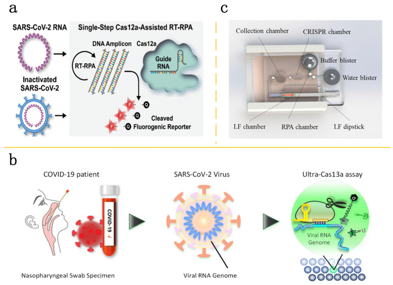
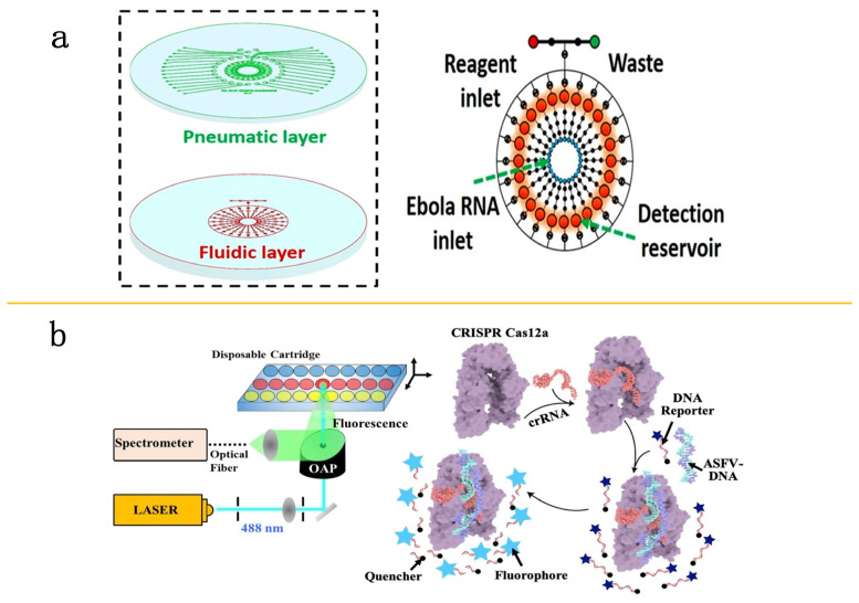
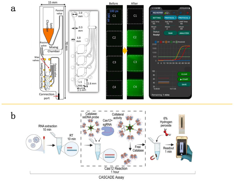
Within the fields of infectious disease diagnostics, microfluidic-based integrated technology systems have become a vital technology in enhancing the rapidity, accuracy, and portability of pathogen detection. These systems synergize microfluidic techniques with advanced molecular biology methods, including reverse transcription polymerase chain reaction (RT-PCR), loop-mediated isothermal amplification (LAMP), and clustered regularly interspaced short palindromic repeats (CRISPR), have been successfully used to identify a diverse array of pathogens, including COVID-19, Ebola, Zika, and dengue fever. This review outlines the advances in pathogen detection, attributing them to the integration of microfluidic technology with traditional molecular biology methods and smartphone- and paper-based diagnostic assays. The cutting-edge diagnostic technologies are of critical importance for disease prevention and epidemic surveillance. Looking ahead, research is expected to focus on increasing detection sensitivity, streamlining testing processes, reducing costs, and enhancing the capability for remote data sharing. These improvements aim to achieve broader coverage and quicker response mechanisms, thereby constructing a more robust defense for global public health security.

Keywords: high throughput; infectious disease; microfluidic system; nucleic acid assay; rapid diagnosis.


Conflict of interest statement

The authors declare no conflicts of interest.

Figures

Figure 7
Figure 7
Paper-based POC microfluidic detection. (a) Schematic of HC-FIA assay. Reproduced with permission [77]. Copyright 2020 Springer Nature. (b) Multiplexed paper devices. Exploded diagram of the compacted device (left) and blueprints of the multi-analyte paper device (right). Reproduced with permission [79]. Copyright 2017 Springer Nature.
Figure 1
Figure 1
Microfluidic system combined with PCR technology. (a) Image of a PF-PCR chip (dimensions: 14 × 26 × 4 mm) (left) accompanied by cross-sectional scanning electron microscopy (SEM) images of PNA (middle). Amplification curves and temperature profiles of lambda DNA (λ-DNA) during a 40-cycle, two-step nano-plasmonic PCR ranging from 65 to 95°, covering 264 s, based on varying initial concentrations of λ-DNA (right). Reproduced with permission [30]. Copyright 2021 American Chemical Society. (b) The fast quantitative RT-qPCR microfluidic setup (left) and the schematic representation of fluid dynamics on a single-use microfluidic chip (right). Reproduced with permission [31]. Copyright 2020 Elsevier Ltd. (c) Flowchart for reanalysis of clinical samples (SARS-CoV-2-positive and -negative). Reproduced with permission [35]. Copyright 2020 MDPI, Basel, Switzerland.
Figure 2
Figure 2
Application of isothermal amplification technique in microfluidic testing. (a) Schematic representation of the LAMP reaction chamber in microfluidic chip: (i) disassembled view; (ii) overhead view; (iii) lateral view. Reproduced with permission [44]. Copyright 2021 Elsevier B.V. (b) The process of the mRT-LAMP-LFB assay consists of four main steps: collecting the sample (3 min), performing rapid RNA extraction (15 min), conducting the mRT-LAMP reaction (40 min), and reporting the results (less than 2 min). This entire RT-LAMP-LFB diagnostic test can be completed in under 60 min. Reproduced with permission [45]. Copyright 2020 Elsevier B.V. (c) Schematic of the visual readout of the LAMP product using a lateral flow assay. During amplification, molecules of biotin and FITC are incorporated into the LAMP product. A test line (T) becomes visible on the assay strip only if the LAMP product is recognized by GNP-Anti-FITC-Ab. Meanwhile, a control line (C) serves as an internal check to confirm the assay’s validity. Reproduced with permission [46]. Copyright 2022 Springer Nature.
Figure 3
Figure 3
The detection technology of isothermal amplification combined with microfluidics. (a) Workflow of the paper-based RT-LAMP assay. Samples spiked with ZIKV were placed onto the loading zone of a paper-based microfluidic device and moved through the channel by capillary action. The circular end (detection zone) of the paper was then cut off. Following this, the RT-LAMP reaction mix was applied to it and the sample was heated on a hot plate at 68 °C for up to 40 min to facilitate amplification. Reproduced with permission [53]. Copyright 2018 Springer Nature. (b) Diagrammatic illustration of the RT-LAMP paper chip designed for concurrent amplification (left) and detection of various viral RNAs (right). Reproduced with permission [54]. Copyright 2019 Elsevier B.V.
Figure 4
Figure 4
Combination of CRISPR-Cas with microfluidic system. (a) A single-step CRISPR/Cas12a-assisted reverse transcription recombinase polymerase amplification (RT-RPA) assay can detect SARS-CoV-2 RNA and inactivated SARS-CoV-2 virus. This method converts RNA targets into DNA amplicons, which then trigger the Cas12a-based cleavage of fluorogenic reporters in one streamlined process. Reproduced with permission [61]. Copyright 2021 Wiley-VCH GmbH. (b) As designed, crRNAs recognize target regions within a viral RNA genome in a droplet, and this interaction initiates the trans-cleavage of quenched fluorescent reporters. Consequently, the droplet “lights up”, signaling the presence of the target of interest. Reproduced with permission [64]. Copyright 2020 American Chemical Society. (c) The microfluidic chip is paired with a heater case powered by a hand warmer, which generates chemical heat. LF stands for lateral flow. Reproduced with permission [65]. Copyright 2021 Elsevier B.V.
Figure 5
Figure 5
Application of CRISPR-Cas technology in microfluidic detection. (a) An automated POC system for EBOV RNA testing. Ebola target RNA is introduced into the detection reservoir, where it undergoes a reaction with Cas13a-crRNA. Reproduced with permission [67]. Copyright 2019 American Chemical Society. (b) Schematic representation of the fluorescence sensing and the CRISPR/Cas12a detection mechanism. CRISPR/Cas12a interacts with crRNA and DNA, forming the Cas12a/crRNA complex, which then cleaves a single-stranded DNA probe to facilitate fluorescence detection. Reproduced with permission [68]. Copyright 2020 Elsevier B.V.
Figure 6
Figure 6
Microfluidic devices combined with smartphones. (a) Schematic representation of the smartphone-based microfluidic chip. The microfluidic chip features several components: a lysis chamber, a mixing chamber, reaction chambers, along with passive and wax valves (left). Chambers C2, C3, and C4 are pre-prepared with SARS-CoV-2 specific primers, whereas Chamber C1 serves as a negative control (middle). The real-time fluorescence signals and temperature profiles from each heater are transmitted to and displayed on a smartphone (right). Reproduced with permission [70]. Copyright 2021 Elsevier B.V. (b) Workflow of the cascade assay combined with smartphones. Viral RNA is isolated from swab samples and then reverse transcribed. It is incorporated into a Cas12 assay, which includes a catalase single-stranded DNA (CD) probe and oxygen. This setup produces a signal through the formation of oxygen bubbles, detectable in a microfluidic channel using a smartphone app. Reproduced with permission [71]. Copyright 2021 Wiley-VCH GmbH.

Similar articles

Cited by

References

    1. Janikddfghx E., Ceremuga M., Niemcewicz M., Bijak M. Dangerous pathogens as a potential problem for public health. Medicina. 2020;56:591. doi: 10.3390/medicina56110591. - DOI - PMC - PubMed
    1. Baker R.E., Mahmud A.S., Miller I.F., Rajeev M., Rasambainarivo F., Rice B.L., Takahashi S., Tatem A.J., Wagner C.E., Wang L.F., et al. Infectious disease in an era of global change. Nat. Rev. Microbiol. 2022;20:193–205. doi: 10.1038/s41579-021-00639-z. - DOI - PMC - PubMed
    1. Onder G., Rezza G., Brusaferro S. Case-Fatality Rate and Characteristics of Patients Dying in Relation to COVID-19 in Italy. JAMA. 2022;323:1775–1776. doi: 10.1001/jama.2020.4683. - DOI - PubMed
    1. Plourde A.R., Bloch E.M. A literature review of zika virus. Emerg. Infect. Dis. 2016;22:1185–1192. doi: 10.3201/eid2207.151990. - DOI - PMC - PubMed
    1. Santiago G.A., Vázquez J., Courtney S., Matías K.Y., Andersen L.E., Colón C., Butler A.E., Roulo R., Bowzard J., Villanueva J.M., et al. Performance of the Trioplex real-time RT-PCR assay for detection of Zika, dengue, and chikungunya viruses. Nat. Commun. 2018;9:1391. doi: 10.1038/s41467-018-03772-1. - DOI - PMC - PubMed

MeSH terms

Supplementary concepts

LinkOut - more resources