. 2024 May 24;29(11):2469.
doi: 10.3390/molecules29112469.

The Application of Ultrasmall Gold Nanoparticles (2 nm) Functionalized with Doxorubicin in Three-Dimensional Normal and Glioblastoma Organoid Models of the Blood-Brain Barrier

Affiliations

The Application of Ultrasmall Gold Nanoparticles (2 nm) Functionalized with Doxorubicin in Three-Dimensional Normal and Glioblastoma Organoid Models of the Blood-Brain Barrier

Kathrin Kostka et al. Molecules. .

Abstract

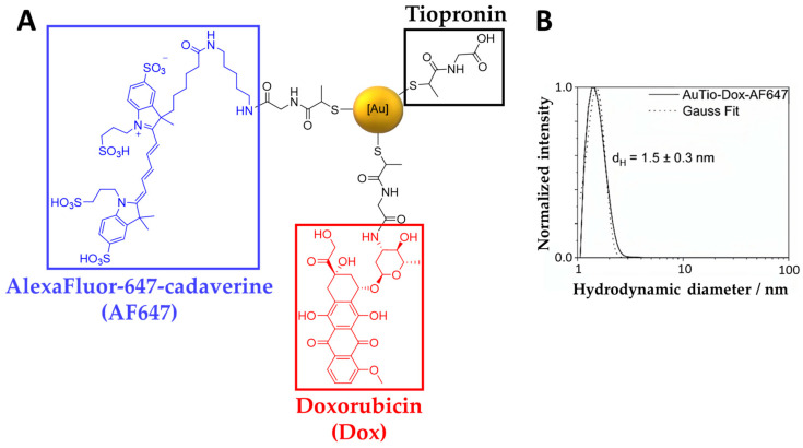
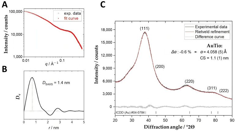
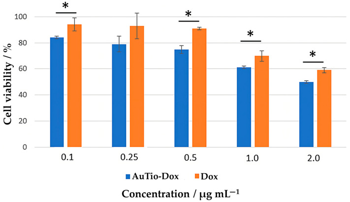
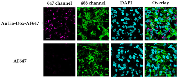
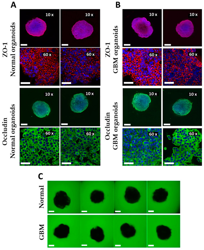
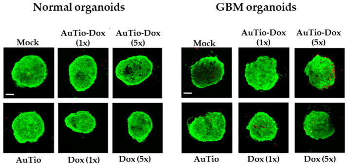
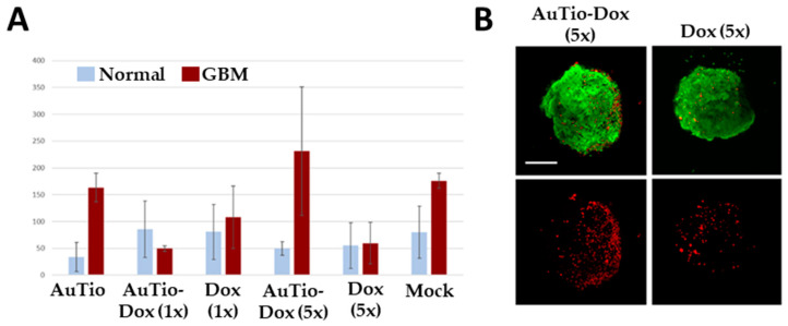
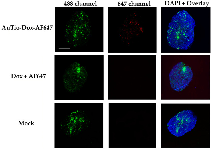
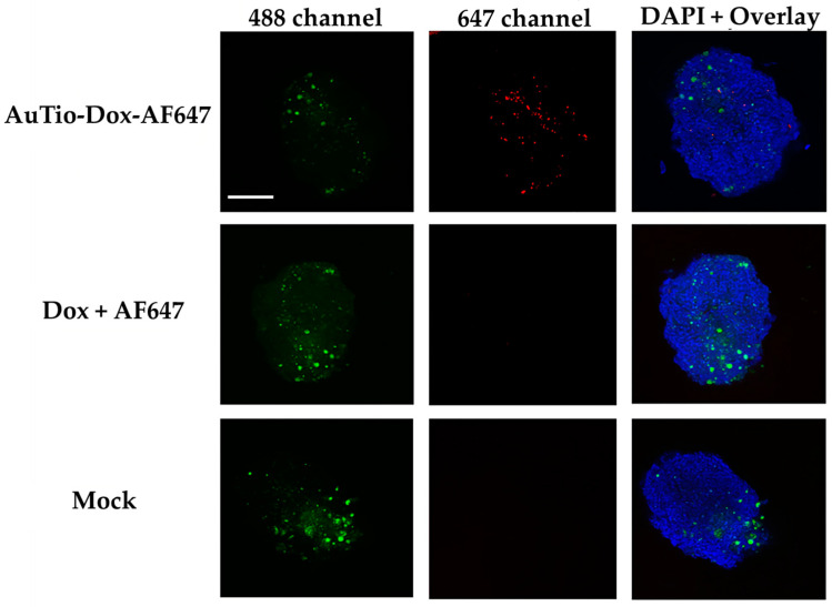
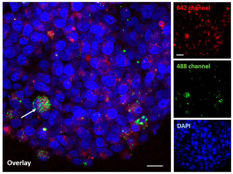
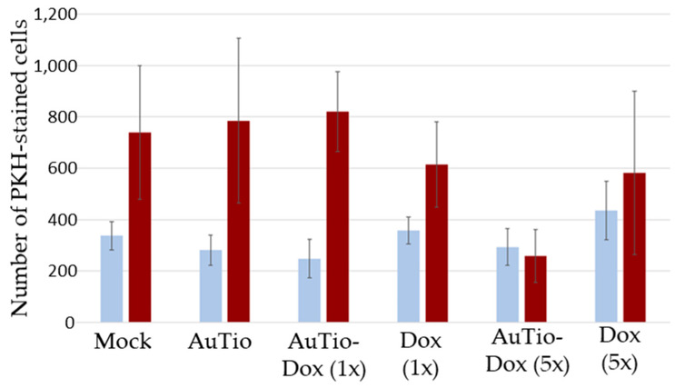
Among brain tumors, glioblastoma (GBM) is very challenging to treat as chemotherapeutic drugs can only penetrate the brain to a limited extent due to the blood-brain barrier (BBB). Nanoparticles can be an attractive solution for the treatment of GBM as they can transport drugs across the BBB into the tumor. In this study, normal and GBM organoids comprising six brain cell types were developed and applied to study the uptake, BBB penetration, distribution, and efficacy of fluorescent, ultrasmall gold nanoparticles (AuTio-Dox-AF647s) conjugated with doxorubicin (Dox) and AlexaFluor-647-cadaverine (AF647) by confocal laser scanning microscopy (CLSM), using a mixture of dissolved doxorubicin and fluorescent AF647 molecules as a control. It was shown that the nanoparticles could easily penetrate the BBB and were found in normal and GBM organoids, while the dissolved Dox and AF647 molecules alone were unable to penetrate the BBB. Flow cytometry showed a reduction in glioblastoma cells after treatment with AuTio-Dox nanoparticles, as well as a higher uptake of these nanoparticles by GBM cells in the GBM model compared to astrocytes in the normal cell organoids. In summary, our results show that ultrasmall gold nanoparticles can serve as suitable carriers for the delivery of drugs into organoids to study BBB function.

Keywords: blood–brain barrier; doxorubicin; drug delivery; glioblastoma; gold; nanoparticles; organoids.


Conflict of interest statement

The authors declare no conflicts of interest.

Figures

Figure 1
Figure 1
The chemical structures (shown schematically) of the ultrasmall gold nanoparticles carrying the fluorescent dye AF647 (left, blue), the cytostatic agent Dox (bottom, red), and the linker tiopronin (right, black) (A). Hydrodynamic particle size distribution as shown by DCS (B).
Figure 2
Figure 2
The results of a SAXS assessment of water-dispersed AuTio-Dox ultrasmall gold nanoparticles with the experimental data I(q) and model fit (A) and the volume-weighed particle size distribution Dv with the determined diameter DSAXS (B). A representative X-ray powder diffractogram (C) with a Rietveld refinement (Rwp = 2.0) of AuTio nanoparticles with calculated lattice parameters (a) and crystallite size (CS). Numbers in parentheses indicate the Miller indices of the diffraction peaks of gold.
Figure 3
Figure 3
MTT assay of T98GBM cells incubated with AuTio-Dox nanoparticles (blue) and Dox molecules alone (orange) as a function of doxorubicin concentration. * = Significance: * p ≤ 0.1.
Figure 4
Figure 4
CLSM images of T98GBM cells incubated for 24 h with either fluorescent ultrasmall gold nanoparticles (AuTio-Dox-AF647s) or AF647 molecules alone. AF647 (magenta; AF647 channel), actin staining with AlexaFluor-488 phalloidin (green; AF488 channel), nuclear staining with DAPI (blue), and an overlay of all channels. Scale bars: 10 µm. The AuTio-Dox-AF647 nanoparticles can be detected as red, fluorescent dots in the cells (first row), whereas no fluorescence is visible after treatment with the AF647 molecules alone (second row).
Figure 5
Figure 5
Characterization of normal and GBM organoids. Organoids were stained with either ZO-1 or occludin, and the nuclei were stained with DAPI for immunostaining ((A) normal organoids and (B) GBM organoids). A higher magnification (×60) was used to visualize the tight junctions (network structures) between endothelial cells. Scale bars: 200 μm (×10) and 50 μm (×60). Permeability was determined with fluorescent dextran (green) after 30 min of incubation (C). The BBB was intact, and no fluorescent signal was visible inside the organoids. Scale bars: 200 μm.
Figure 6
Figure 6
Live/dead assay. Living cells are shown in green, and dead cells are shown in red. Normal and GBM organoids after incubation with AuTio-Dox nanoparticles and doxorubicin molecules alone; (1×) corresponds to a normal concentration and (5×) corresponds to a five times higher concentration of AuTio-Dox (5×) or doxorubicin (5×), respectively. Scale bars: 200 µm.
Figure 7
Figure 7
Live/dead assay. Average number of red dead cells calculated from five organoids (A). Determination of average number of red cells quantified inside brain organoids after uptake of AuTio-Dox nanoparticles and doxorubicin molecules alone after 24 h of incubation. Number of red cells was calculated and averaged from n = 5 organoids using FIJI. Representative images are shown for AuTio-Dox (5×) and Dox (5×) from CLSM (B). Scale bars: 200 µm.
Figure 8
Figure 8
Uptake studies of the AuTio-Dox-AF647 nanoparticles and a mixture of dissolved doxorubicin and AF647 in normal organoids after 24 h of incubation. Astrocytes were fluorescently labeled with PKH-CellTracker (green; 488 channel). The AuTio-Dox-AF647 nanoparticles showed red fluorescence (647 channel; first row) in the organoids, while the mixture of doxorubicin and AF647 molecules showed no fluorescence (second row). Scale bars: 200 µm.
Figure 9
Figure 9
Uptake studies of AuTio-Dox-AF647s and dissolved doxorubicin and AF647 in GBM organoids after 24 h of incubation. U87-MG cells were fluorescently labeled with PKH-CellTracker (green; 488 channel). The nanoparticles showed red fluorescence. The AuTio-Dox-AF647 nanoparticles entered the organoid (red dots; 647 channel), whereas the doxorubicin and AF647 molecules alone showed no fluorescence (second row). Scale bars: 200 µm.
Figure 10
Figure 10
The uptake of AuTio-Dox-AF647s in a GBM organoid after 24 h at a higher magnification. The nanoparticles show a red fluorescence (647 channel), and the GBM organoid shows a green fluorescence (with arrow; labeled with PKH-CellTracker; green; 488 channel). A good distribution of the AuTio-Dox-AF647 nanoparticles in the organoid can be visualized. Scale bars: 10 µm.
Figure 11
Figure 11
Number of PKH-CellTracker-stained cells (astrocytes in normal organoids and U87-MG cells in GBM organoids) quantified from five organoids using CLSM images of normal and GBM organoids. The determination of the number of PKH-CellTracker-stained cells quantified from inside the BBB organoids after the uptake of ultrasmall gold nanoparticles and doxorubicin molecules after 24 h of incubation. The average was calculated using the software FIJI.

Similar articles

Cited by

References

    1. Alifieris C., Trafalis D.T. Glioblastoma multiforme: Pathogenesis and treatment. Pharmacol. Ther. 2015;152:63–82. doi: 10.1016/j.pharmthera.2015.05.005. - DOI - PubMed
    1. Taylor O.G., Brzozowski J.S., Skelding K.A. Glioblastoma Multiforme: An Overview of Emerging Therapeutic Targets. Front. Oncol. 2019;9:963. doi: 10.3389/fonc.2019.00963. - DOI - PMC - PubMed
    1. Alexander B.M., Cloughesy T.F. Adult Glioblastoma. J. Clin. Oncol. 2017;35:2402–2409. doi: 10.1200/JCO.2017.73.0119. - DOI - PubMed
    1. Paolillo M., Boselli C., Schinelli S. Glioblastoma under Siege: An Overview of Current Therapeutic Strategies. Brain Sci. 2018;8:15. doi: 10.3390/brainsci8010015. - DOI - PMC - PubMed
    1. Caragher S., Chalmers A.J., Gomez-Roman N. Glioblastoma’s Next Top Model: Novel Culture Systems for Brain Cancer Radiotherapy Research. Cancers. 2019;11:44. doi: 10.3390/cancers11010044. - DOI - PMC - PubMed

LinkOut - more resources