. 2024 May 30;29(11):2579.
doi: 10.3390/molecules29112579.

Structural Analysis of the Large Stokes Shift Red Fluorescent Protein tKeima

Affiliations

Structural Analysis of the Large Stokes Shift Red Fluorescent Protein tKeima

Ki Hyun Nam et al. Molecules. .

Abstract

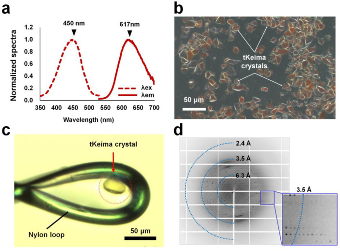
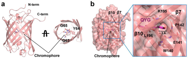
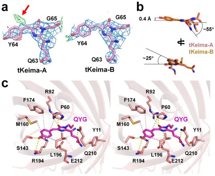
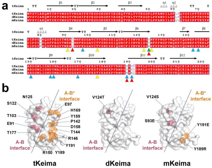
The Keima family comprises large Stokes shift fluorescent proteins that are useful for dual-color fluorescence cross-correlation spectroscopy and multicolor imaging. The tKeima is a tetrameric large Stokes shift fluorescent protein and serves as the ancestor fluorescent protein for both dKeima and mKeima. The spectroscopic properties of tKeima have been previously reported; however, its structural basis and molecular properties have not yet been elucidated. In this study, we present the crystallographic results of the large Stokes shift fluorescent protein tKeima. The purified tKeima protein spontaneously crystallized after purification without further crystallization. The crystal structure of tKeima was determined at 3.0 Å resolution, revealing a β-barrel fold containing the Gln-Tyr-Gly chromophores mainly with cis-conformation. The tetrameric interfaces of tKeima were stabilized by numerous hydrogen bonds and salt-bridge interactions. These key residues distinguish the substituted residues in dKeima and mKeima. The key structure-based residues involved in the tetramer formation of tKeima provide insights into the generation of a new type of monomeric mKeima. This structural analysis expands our knowledge of the Keima family and provides insights into its protein engineering.

Keywords: Keima; fluorescent protein; large Stoke shift; structure; tKeima.


Conflict of interest statement

The authors declare no conflicts of interest.

Figures

Figure 1
Figure 1
Spectroscopic and crystallographic analysis of tKeima. (a) Spectroscopic analysis of purified tKeima. The maximum excitation and emission peaks of tKeima were at 450 and 617 nm, respectively. (b) Microscopic view of spontaneously grown tKeima crystals. (c) The tKeima crystal was used for diffraction data collection. (d) Diffraction pattern of the spontaneously grown tKeima crystal.
Figure 2
Figure 2
Crystal structure of tKeima. (a) Cartoon representation of the tKeima monomer. The chromophore consisting of the Gln-Tyr-Gly tripeptide located at the center of the β-barrel. (b) Surface structure of tKeima. A solvent-accessible hole was observed between β7 and β10 strands of the β-barrel.
Figure 3
Figure 3
The tKeima chromophore and its environment. (a) Electron density maps of 2mFo-DFc (blue, 1.0 σ) and mFo-DFc (green, 3.0 σ and red, −3.0 σ) of the tKeima chromophore. The trans-conformation of the chromophore is indicated by the red arrow. (b) Analysis of the tKeima chromophore. (c) Stereo view of the tKeima chromophore and its interacting residues.
Figure 4
Figure 4
Structural analysis of tetrameric tKeima. (a) Tetrameric arrangement of tKeima with 222 symmetry. (b) B-factor putty representation of tKeima. (c) Close-up and (d) open-book view of the interaction residues of interface A–B of tKeima. Hydrogen bonds and salt bridges are indicated by the dotted line. (e) Close-up and (f) open-book view of the interaction residues of interfaces A–B* of tKeima. (g) Structural comparison of the tetrameric assembly of tKeima, DsRed (PDB code: 1GGX), AdRed (6AA7), ZsYellow (6LOF), and DendFP (7DIG).
Figure 5
Figure 5
A comparison of the amino acids and structures of the Keima family. (a) Structure-based sequence alignment of tKeima (UniProt: Q1JV72), dKeima (Q1JV71), and mKeima (Q1JV70). The residues involved in the oligomer states of tKeima are indicated using yellow (A–B interface) and blue (A–B* interface) triangles. The mutation sites located on the surface of dKeima and mKeima are indicated by green and red triangles, respectively. (b) Surface structure of key residue involved in the tetrameric interface of tKeima and mutation sites located on the surface of dKeima and mKeima.

Similar articles

Cited by

References

    1. Bajar B.T., Wang E.S., Zhang S., Lin M.Z., Chu J. A Guide to Fluorescent Protein FRET Pairs. Sensors. 2016;16:1488. doi: 10.3390/s16091488. - DOI - PMC - PubMed
    1. Bizzarri R., Arcangeli C., Arosio D., Ricci F., Faraci P., Cardarelli F., Beltram F. Development of a Novel GFP-based Ratiometric Excitation and Emission pH Indicator for Intracellular Studies. Biophys. J. 2006;90:3300–3314. doi: 10.1529/biophysj.105.074708. - DOI - PMC - PubMed
    1. Frommer W.B., Davidson M.W., Campbell R.E. Genetically encoded biosensors based on engineered fluorescent proteins. Chem. Soc. Rev. 2009;38:2833. doi: 10.1039/b907749a. - DOI - PMC - PubMed
    1. Benaissa H., Ounoughi K., Aujard I., Fischer E., Goïame R., Nguyen J., Tebo A.G., Li C., Le Saux T., Bertolin G., et al. Engineering of a fluorescent chemogenetic reporter with tunable color for advanced live-cell imaging. Nat. Commun. 2021;12:6989. doi: 10.1038/s41467-021-27334-0. - DOI - PMC - PubMed
    1. Miura Y., Senoo A., Doura T., Kiyonaka S. Chemogenetics of cell surface receptors: Beyond genetic and pharmacological approaches. RSC Chem. Biol. 2022;3:269–287. doi: 10.1039/d1cb00195g. - DOI - PMC - PubMed

Substances

LinkOut - more resources