

Quality Studies on Cynometra iripa Leaf and Bark as Herbal Medicines
- PMID: 38893505
- PMCID: PMC11173719
- DOI: 10.3390/molecules29112629
Quality Studies on Cynometra iripa Leaf and Bark as Herbal Medicines
Abstract
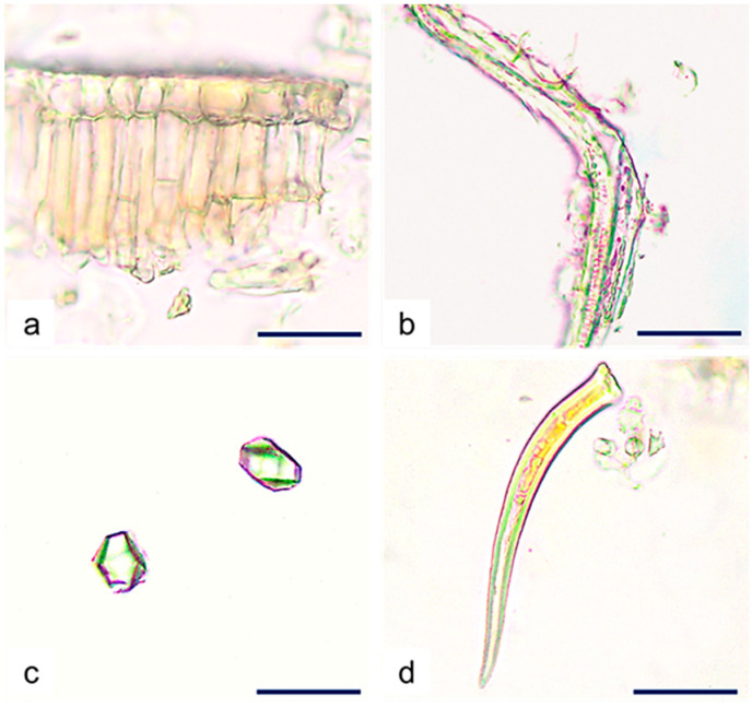
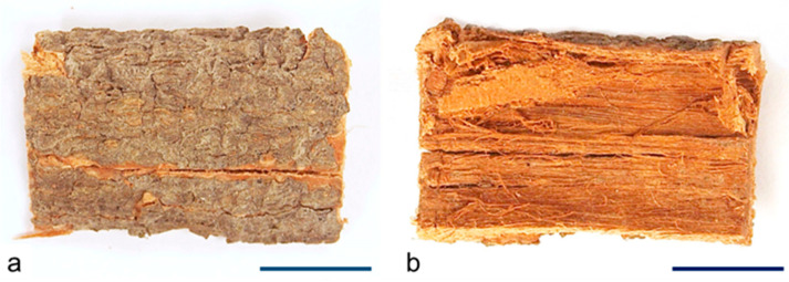
Cynometra iripa Kostel. is a Fabaceae species of mangrove used in traditional Ayurvedic medicine for treating inflammatory conditions. The present study aims to establish monographic botanical and chemical quality criteria for C. iripa leaf and bark as herbal substances and to evaluate their in vitro antioxidant potential. Macroscopic and microscopic qualitative and quantitative analyses, chemical LC-UV/DAD-ESI/MS profiling, and the quantification of key chemical classes were performed. Antioxidant activity was evaluated by DPPH and FRAP assays. Macroscopically, the leaf is asymmetrical with an emarginated apex and cuneate base. Microscopically, it shows features such as two-layered adaxial palisade parenchyma, vascular bundles surrounded by 3-6 layers of sclerenchyma, prismatic calcium oxalate crystals (5.89 ± 1.32 μm) along the fibers, paracytic stomata only on the abaxial epidermis (stomatal index-20.15), and non-glandular trichomes only on petiolules. The microscopic features of the bark include a broad cortex with large lignified sclereids, prismatic calcium oxalate crystals (8.24 ± 1.57 μm), and secondary phloem with distinct 2-5 seriated medullary rays without crystals. Chemical profile analysis revealed that phenolic derivatives, mainly condensed tannins and flavonoids, are the main classes identified. A total of 22 marker compounds were tentatively identified in both plant parts. The major compounds identified in the leaf were quercetin-3-O-glucoside and taxifolin pentoside and in the bark were B-type dimeric proanthocyanidins and taxifolin 3-O-rhamnoside. The total phenolics content was higher in the leaf (1521 ± 4.71 mg GAE/g dry weight), while the total flavonoids and condensed tannins content were higher in the bark (82 ± 0.58 mg CE/g and 1021 ± 5.51 mg CCE/g dry weight, respectively). A total of 70% of the hydroethanolic extracts of leaf and bark showed higher antioxidant activity than the ascorbic acid and concentration-dependent scavenging activity in the DPPH assay (IC50 23.95 ± 0.93 and 23.63 ± 1.37 µg/mL, respectively). A positive and statistically significant (p < 0.05) correlation between the phenol content and antioxidant activity was found. The results obtained will provide important clues for the quality control criteria of C. iripa leaf and bark, as well as for the knowledge of their pharmacological potential as possible anti-inflammatory agents with antioxidant activity.
Keywords: Cynometra iripa; antioxidant; chemical profile; herbal substance; light microscopy; macroscopic analysis; mangrove species; quality control.
Conflict of interest statement
The authors confirm that the content of this article has no conflicts of interest.
Figures


References
-
- The IUCN Red List of Threatened Species. Cynometra iripa. 2023. [(accessed on 5 January 2023)]. Available online: https://www.iucnredlist.org/species/178829/7619971.
-
- Duke N.C. Mangrove floristics and biogeography. In: Robertson A.I., Alongi D.M., editors. Tropical Mangrove Ecosystems. American Geophysical Union; Washington, DC, USA: 1992. pp. 63–100. - DOI
-
- Polidoro B.A., Carpenter K.E., Collins L., Duke N.C., Ellison A.M., Ellison J.C., Farnsworth E.J., Fernando E.S., Kathiresan K., Koedam N.E., et al. The loss of species: Mangrove extinction risk and geographic areas of global concern. PLoS ONE. 2010;5:e10095. doi: 10.1371/journal.pone.0010095. - DOI - PMC - PubMed
-
- Bruneau A., Forest F., Herendeen P.S., Klitgaard B.B., Lewis G.P. Phylogenetic relationships in the Caesalpinioideae (Leguminosae) as inferred from chloroplast trnL intron sequences. Syst. Bot. 2001;26:487–514. doi: 10.1043/0363-6445-26.3.487. - DOI
-
- Bruneau A., Mercure M., Lewis G.P., Herendeen P.S. Phylogenetic patterns and diversification in the Caesalpinioid legumes. Botany. 2008;86:697–718. doi: 10.1139/B08-058. - DOI
MeSH terms
Substances
Grants and funding
LinkOut - more resources
Full Text Sources
Medical
Miscellaneous
