Review
. 2024 Jun 3;29(11):2635.
doi: 10.3390/molecules29112635.

Decoding the κ Opioid Receptor (KOR): Advancements in Structural Understanding and Implications for Opioid Analgesic Development

Affiliations
Review

Decoding the κ Opioid Receptor (KOR): Advancements in Structural Understanding and Implications for Opioid Analgesic Development

Zoe Li et al. Molecules. .

Abstract

The opioid crisis in the United States is a significant public health issue, with a nearly threefold increase in opioid-related fatalities between 1999 and 2014. In response to this crisis, society has made numerous efforts to mitigate its impact. Recent advancements in understanding the structural intricacies of the κ opioid receptor (KOR) have improved our knowledge of how opioids interact with their receptors, triggering downstream signaling pathways that lead to pain relief. This review concentrates on the KOR, offering crucial structural insights into the binding mechanisms of both agonists and antagonists to the receptor. Through comparative analysis of the atomic details of the binding site, distinct interactions specific to agonists and antagonists have been identified. These insights not only enhance our understanding of ligand binding mechanisms but also shed light on potential pathways for developing new opioid analgesics with an improved risk-benefit profile.

Keywords: agonist; antagonist; binding; ligand; mechanism; opioid; receptor; structure.


Conflict of interest statement

The authors declare no conflicts of interest.

Figures

Figure 1
Figure 1
Sequence alignment result. The upper panel shows the sequence alignment of KOR, MOR, and DOR. The lower panel shows the percent identity matrix of the sequence alignment. Amino acids common to all subtypes are highlighted in dark purple, while those shared only by two subtypes are marked in light purple.
Figure 2
Figure 2
Alignment of five KOR structures, with one structure bound with an antagonist (depicted in blue) and the remaining four structures bound with agonists (depicted in white). The right panel shows an enlarged view, showcasing the alignment of all ligands, both the agonists and antagonist.
Figure 3
Figure 3
Two-dimensional structures of the ligands.
Figure 4
Figure 4
The ligand binding poses in KOR. The upper panel displays the antagonist binding pose and the lower panel showcases the agonist binding pose.
Figure 5
Figure 5
The momSalB-bound (colored in beige) KOR (PDB ID: 8DZP, colored in white) is aligned with agonist-bound MOR (PDB ID: 8EF6, colored in pink) and agonist-bound DOR (PDB ID: 6PT3, colored in light blue). The V1082.53 of KOR and the corresponding alanine in both DOR and MOR is represented in stick and circled in red.
Figure 6
Figure 6
(a) Venn diagram showing the common and distinct residues found within 3.5 Å for antagonist (depicted by the blue circles) and agonist (depicted by the red circles). (b) Residues within 3.5 Å for antagonist JDTic (PDB ID: 6VI4): the residue unique to antagonist is colored in light blue, residue common to both agonist and antagonist is colored in light green, and the residue that is partially shared between agonist and antagonist is colored in light purple. (cf) Residues within 3.5 Å for agonist MP1140 (PDB ID: 6B73), nalfurafine (PDB ID: 7YIT), and GR89696 (PDB ID: 8DZR), momSalB (PDB ID: 8DZP) respectively. The residue unique to agonist is colored in light coral, residue common to both agonist and antagonist is colored in light green, and the residue that is partially shared between agonist and antagonist is colored in light purple. All ligands are colored in yellow.
Figure 6
Figure 6
(a) Venn diagram showing the common and distinct residues found within 3.5 Å for antagonist (depicted by the blue circles) and agonist (depicted by the red circles). (b) Residues within 3.5 Å for antagonist JDTic (PDB ID: 6VI4): the residue unique to antagonist is colored in light blue, residue common to both agonist and antagonist is colored in light green, and the residue that is partially shared between agonist and antagonist is colored in light purple. (cf) Residues within 3.5 Å for agonist MP1140 (PDB ID: 6B73), nalfurafine (PDB ID: 7YIT), and GR89696 (PDB ID: 8DZR), momSalB (PDB ID: 8DZP) respectively. The residue unique to agonist is colored in light coral, residue common to both agonist and antagonist is colored in light green, and the residue that is partially shared between agonist and antagonist is colored in light purple. All ligands are colored in yellow.
Figure 7
Figure 7
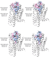
Comparison of the solvent-accessible volume (SAV) in the antagonist-bound structure (SAV depicted in blue) with that in the agonist-bound structures (SAV highlighted in pink).
Figure 8
Figure 8
Comparison of the binding pocket RMSD between the antagonist-bound and agonist-bound structures. The left panel lists RMSD for all pairs of the structures. The right panel gives RMSD for each of the five structures aligned with the other four structures. The average of the four RMSD values for each of the five structures is plotted as a bar, and the corresponding standard deviation is presented by the stick on the bar. Structures of apo KOR were not included in the comparison, as our interest is to examine the difference between agonist-binding pocket and antagonist-binding pocket.

Similar articles

References

    1. Rudd R.A., Seth P., David F., Scholl L. Increases in Drug and Opioid-Involved Overdose Deaths—United States, 2010–2015. MMWR Morb. Mortal. Wkly. Rep. 2016;65:1445–1452. doi: 10.15585/mmwr.mm655051e1. - DOI - PubMed
    1. Rudd R.A., Aleshire N., Zibbell J.E., Gladden R.M. Increases in Drug and Opioid Overdose Deaths--United States, 2000–2014. MMWR Morb. Mortal. Wkly. Rep. 2016;64:1378–1382. doi: 10.15585/mmwr.mm6450a3. - DOI - PubMed
    1. Jones C.M., Baldwin G.T., Manocchio T., White J.O., Mack K.A. Trends in Methadone Distribution for Pain Treatment, Methadone Diversion, and Overdose Deaths—United States, 2002–2014. MMWR Morb. Mortal. Wkly. Rep. 2016;65:667–671. doi: 10.15585/mmwr.mm6526a2. - DOI - PubMed
    1. Peterson A.B., Gladden R.M., Delcher C., Spies E., Garcia-Williams A., Wang Y., Halpin J., Zibbell J., McCarty C.L., DeFiore-Hyrmer J., et al. Increases in Fentanyl-Related Overdose Deaths—Florida and Ohio, 2013–2015. MMWR Morb. Mortal. Wkly. Rep. 2016;65:844–849. doi: 10.15585/mmwr.mm6533a3. - DOI - PubMed
    1. Gladden R.M., Martinez P., Seth P. Fentanyl Law Enforcement Submissions and Increases in Synthetic Opioid-Involved Overdose Deaths—27 States, 2013–2014. MMWR Morb. Mortal. Wkly. Rep. 2016;65:837–843. doi: 10.15585/mmwr.mm6533a2. - DOI - PubMed

LinkOut - more resources