. 2024 Jun 5;29(11):2676.
doi: 10.3390/molecules29112676.

Non-Glycosylated SARS-CoV-2 Omicron BA.5 Receptor Binding Domain (RBD) with a Native-like Conformation Induces a Robust Immune Response with Potent Neutralization in a Mouse Model

Affiliations

Non-Glycosylated SARS-CoV-2 Omicron BA.5 Receptor Binding Domain (RBD) with a Native-like Conformation Induces a Robust Immune Response with Potent Neutralization in a Mouse Model

Rawiwan Wongnak et al. Molecules. .

Abstract

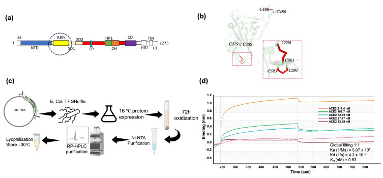
The Omicron BA.5 variant of SARS-CoV-2 is known for its high transmissibility and its capacity to evade immunity provided by vaccine protection against the (original) Wuhan strain. In our prior research, we successfully produced the receptor-binding domain (RBD) of the SARS-CoV-2 spike protein in an E. coli expression system. Extensive biophysical characterization indicated that, even without glycosylation, the RBD maintained native-like conformational and biophysical properties. The current study explores the immunogenicity and neutralization capacity of the E. coli-expressed Omicron BA.5 RBD using a mouse model. Administration of three doses of the RBD without any adjuvant elicited high titer antisera of up to 7.3 × 10⁵ and up to 1.6 × 10⁶ after a booster shot. Immunization with RBD notably enhanced the population of CD44+CD62L+ T cells, indicating the generation of T cell memory. The in vitro assays demonstrated the antisera's protective efficacy through significant inhibition of the interaction between SARS-CoV-2 and its human receptor, ACE2, and through potent neutralization of a pseudovirus. These findings underscore the potential of our E. coli-expressed RBD as a viable vaccine candidate against the Omicron variant of SARS-CoV-2.

Keywords: E. coli-expression system; Omicron SARS-CoV-2; disulfide bonds; immunogenicity; limited proteolysis; molten globule state; native-like state; neutralization; non-glycosylation.


Conflict of interest statement

The authors declare no conflicts of interest.

Figures

Figure 1
Figure 1
SARS-CoV-2 Omicron BA.5 RBD expression and purification in E. coli. (a) Schematics of the sequence location of RBD in the SARS-CoV-2 spike protein. (b) Ribbon model of SARS-CoV-2 RBD with disulfide bond pairing. (c) SARS-CoV-2 Omicron BA.5 RBD expression and purification protocol in E. coli. (d) Binding of the SARS-CoV-2 Omicron BA.5 RBD to the hACE2 using an Octet-N1 Bio-Layer Interferometer. RBD was immobilized on a Ni-NTA sensor chip, and hACE2 was in the mobile phase.
Figure 2
Figure 2
The immunogenicity of E. coli-expressed RBD in a mouse model. (a) Immunization scheme with adjuvant, no adjuvant, and control groups. (b) IgG titer assays by ELISA of RBD-immunized mice with adjuvant (n = 4). Individual mouse IgG titers are represented by circles, and the average titer is presented in bars. (c) IgG titer assays by ELISA of RBD-immunized mice without adjuvant (n = 4). Individual mouse IgG titers are represented by circles, and the average titer is represented with bars. (d) Recognition of mammalian-expressed S1 spike protein by E. coli-expressed RBD antisera (1:1000 dilution). Circles represent the OD at 492 nm of four mice from each group (adjuvant and no adjuvant), indicating the binding of antisera collected in the 9th week after the first administration. Measurements were taken using mammalian-expressed spike (S1) protein or E. coli-expressed RBD as coating antigens for comparison. The average OD value of each group is presented in bars. The analysis revealed no significant difference (p > 0.05) between the S1-coated and RBD-coated results, ‘ns’ stands for not significant.
Figure 3
Figure 3
Effects of E. coli-expressed RBD immunization on T-cell memory assessed through CD marker analysis by flow cytometry. (a) The bar graph illustrates the percentage of CD44+CD62L+ T cells in the control mouse (n = 1) compared with the average percentage of CD44+CD62L+ T cells in two mice (n = 2) from the adjuvant and no adjuvant groups. The individual data are represented by circles. (b) Cluster of differentiation (CD) expression of CD4+ (T-helper cell) surface in the single mouse from each group. (c) Cluster of differentiation (CD) expression of CD8+ (T-cytolytic cell) surface in the single mouse from each group. Note: Due to the absence of multiple data points within the control group (n = 1), no statistical analysis was performed.
Figure 4
Figure 4
hACE2 inhibition assay. (a) Steps of the hACE2 binding inhibition assay using bio-layer interferometry (BLI). RBD was immobilized on the biosensor chip, followed by antisera binding. hACE2 was loaded for the association and dissociation steps assessed in the kinetic buffer. (b) Enlarged figure of hACE2 association and dissociation step. (c) Inhibition of RBD binding to hACE2 by E. coli-expressed RBD-immunized antisera of one mouse from each group; “M” indicates the identity of the mouse.
Figure 5
Figure 5
Pseudovirus neutralization assay. (a) Neutralization (%) of pseudovirus by antisera at 141 days after the first administration of RBD. Neutralization percentages of individual mice (n = 4) from the no adjuvant group are represented by circles, and the average is presented with bars. (b) Pseudovirus neutralization titers (ID50) analysis of a single mouse from the no adjuvant group (M7). The inhibition rate against pseudovirus is plotted against the reciprocal of the antisera dilution. Note: RBD-immunized data (n = 4) showed 100% neutralization with no variance (mean = 100%, standard deviation = 0%), while the single data point in the control group showed 0% neutralization. Due to the lack of variability in the RBD-immunized group, traditional statistical tests such as the t-test were not applicable. Descriptive statistics are provided to illustrate the results.

Similar articles

References

    1. Geng Q., Shi K., Ye G., Zhang W., Aihara H., Li F. Structural Basis for Human Receptor Recognition by SARS-CoV-2 Omicron Variant BA.1. J. Virol. 2022;96:e0024922. doi: 10.1128/jvi.00249-22. - DOI - PMC - PubMed
    1. Tallei T.E., Alhumaid S., AlMusa Z., Fatimawali, Kusumawaty D., Alynbiawi A., Alshukairi A.N., Rabaan A.A. Update on the Omicron Sub-Variants BA.4 and BA.5. Rev. Med. Virol. 2023;33:e2391. doi: 10.1002/rmv.2391. - DOI - PMC - PubMed
    1. Tegally H., Moir M., Everatt J., Giovanetti M., Scheepers C., Wilkinson E., Subramoney K., Makatini Z., Moyo S., Amoako D.G., et al. Emergence of SARS-CoV-2 Omicron Lineages BA.4 and BA.5 in South Africa. Nat. Med. 2022;28:1785–1790. doi: 10.1038/s41591-022-01911-2. - DOI - PMC - PubMed
    1. Tuekprakhon A., Nutalai R., Dijokaite-Guraliuc A., Zhou D., Ginn H.M., Selvaraj M., Liu C., Mentzer A.J., Supasa P., Duyvesteyn H.M.E., et al. Antibody Escape of SARS-CoV-2 Omicron BA.4 and BA.5 from Vaccine and BA.1 Serum. Cell. 2022;185:2422–2433.e13. doi: 10.1016/j.cell.2022.06.005. - DOI - PMC - PubMed
    1. Servellita V., Syed A.M., Morris M.K., Brazer N., Saldhi P., Garcia-Knight M., Sreekumar B., Khalid M.M., Ciling A., Chen P.Y., et al. Neutralizing Immunity in Vaccine Breakthrough Infections from the SARS-CoV-2 Omicron and Delta Variants. Cell. 2022;185:1539. doi: 10.1016/j.cell.2022.03.019. - DOI - PMC - PubMed

MeSH terms

Supplementary concepts