Review
. 2024 May 21;14(11):1064.
doi: 10.3390/diagnostics14111064.

Neuroimaging of the Most Common Meningitis and Encephalitis of Adults: A Narrative Review

Affiliations
Review

Neuroimaging of the Most Common Meningitis and Encephalitis of Adults: A Narrative Review

Teresa Perillo et al. Diagnostics (Basel). .

Abstract

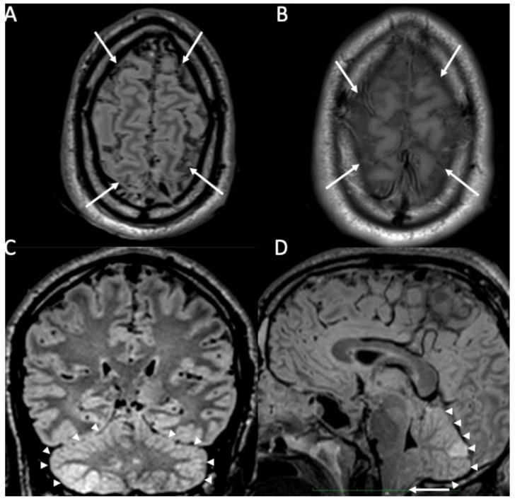
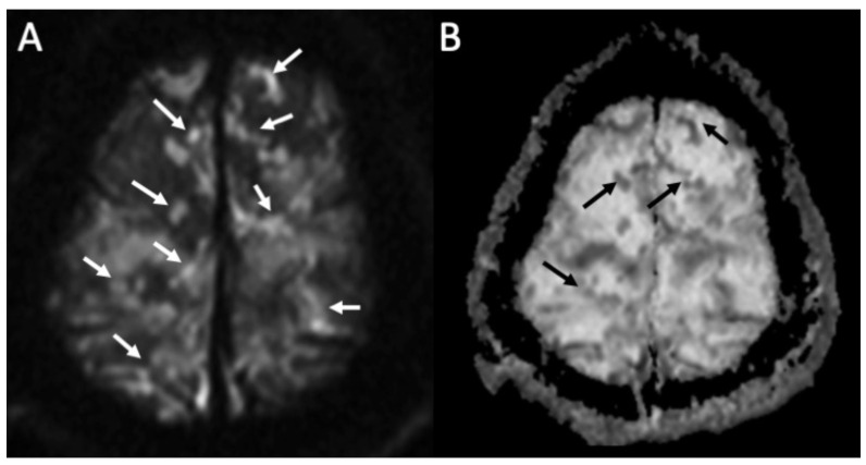
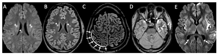
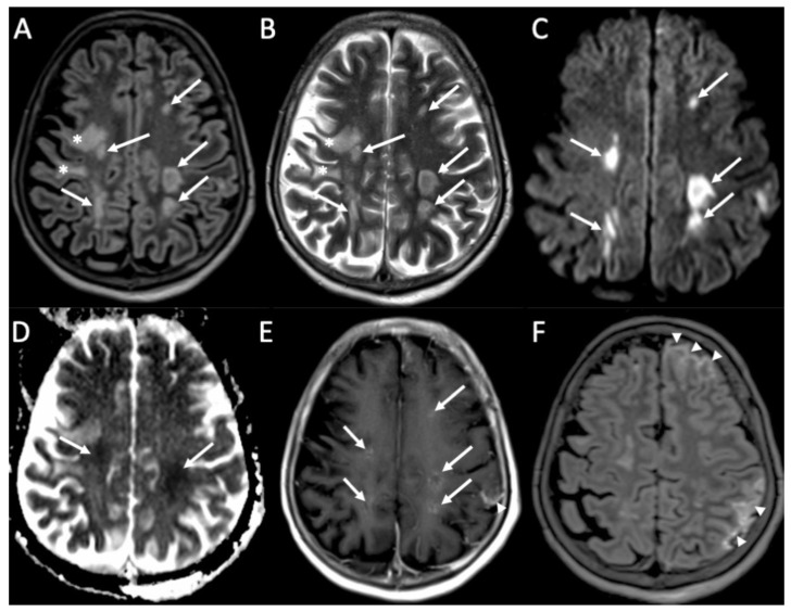
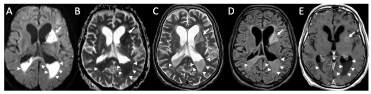
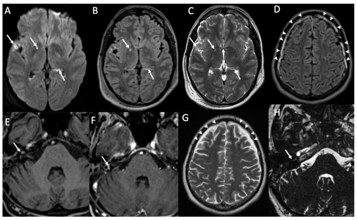
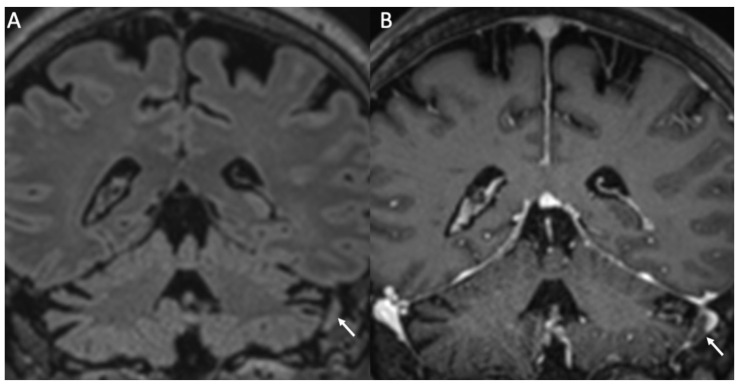
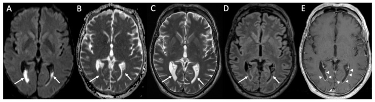
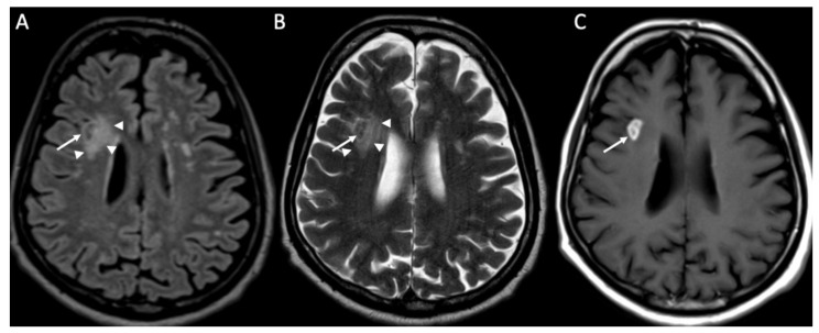
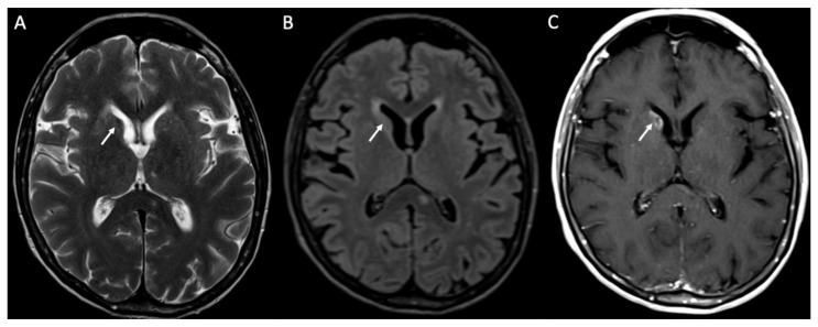
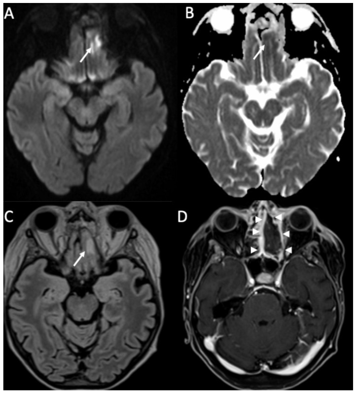
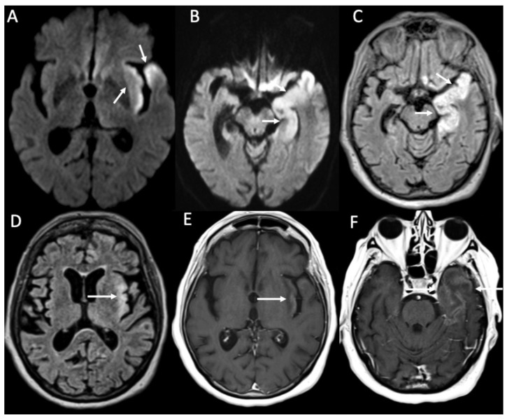
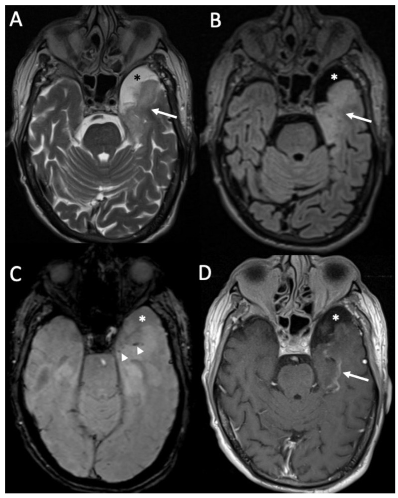
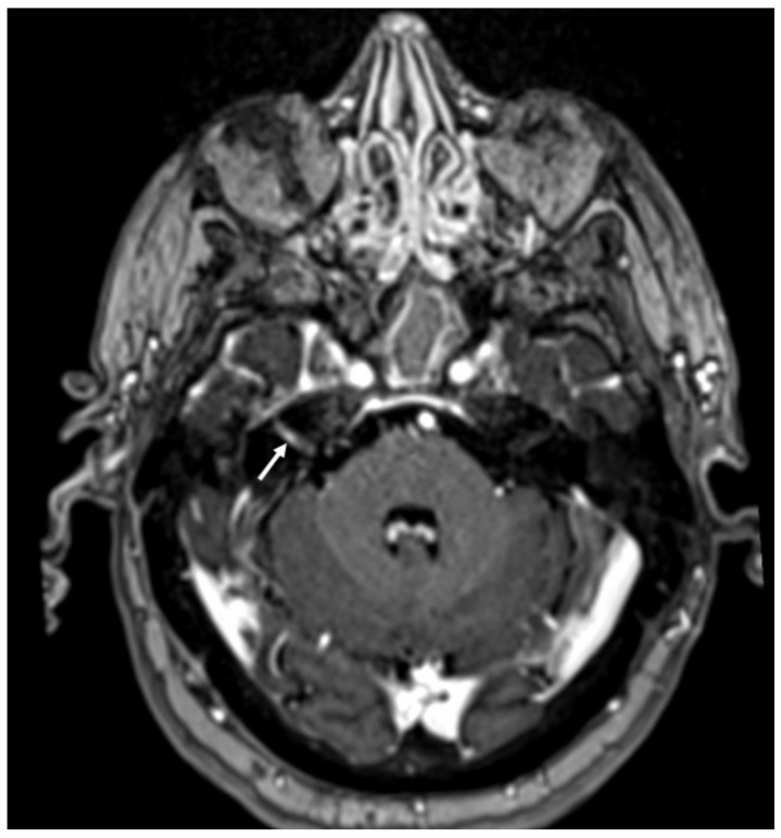
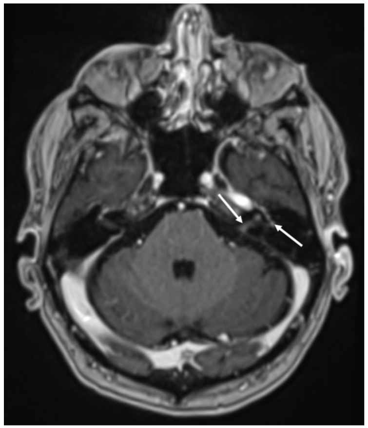
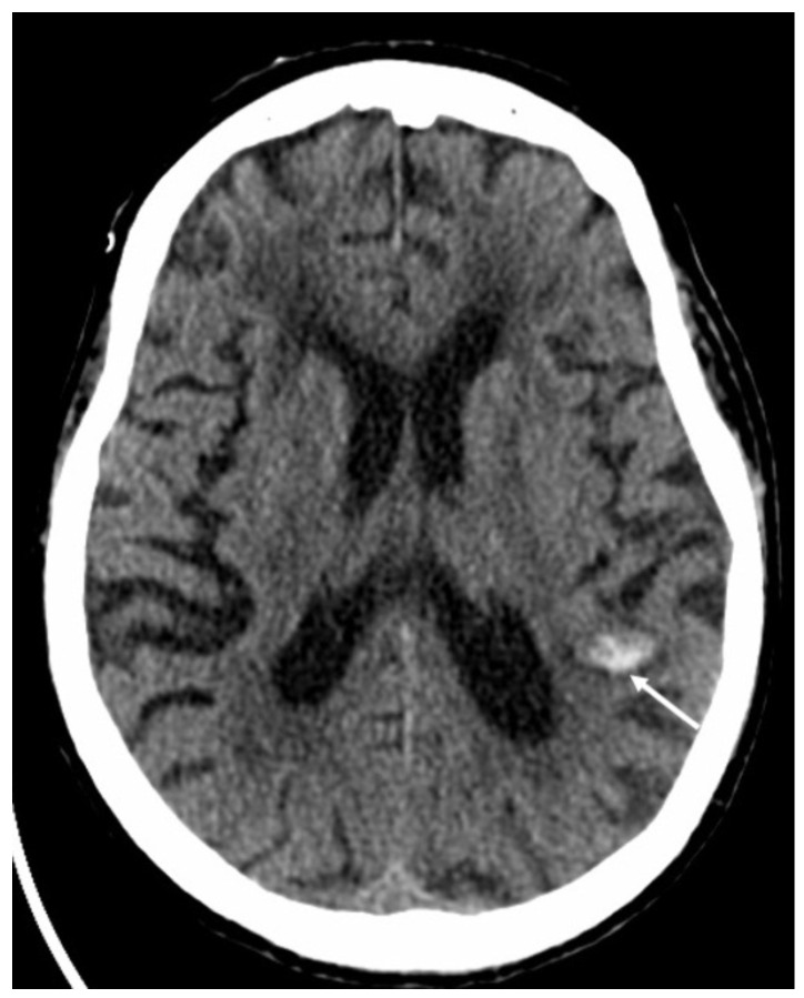
Meningitis is the infection of the meninges, which are connective tissue membranes covering the brain, and it most commonly affects the leptomeninges. Clinically, meningitis may present with fever, neck stiffness, altered mental status, headache, vomiting, and neurological deficits. Encephalitis is an infection of the brain, which usually presents with fever, altered mental status, neurological deficits, and seizure. Meningitis and encephalitis are serious conditions which could also coexist, with high morbidity and mortality, thus requiring prompt diagnosis and treatment. Imaging plays an important role in the clinical management of these conditions, especially Magnetic Resonance Imaging. It is indicated to exclude mimics and evaluate the presence of complications. The aim of this review is to depict imaging findings of the most common meningitis and encephalitis.

Keywords: computed tomography; encephalitis; magnetic resonance imaging; meningitis.


Conflict of interest statement

The authors declare no conflicts of interest.

Figures

Figure 1
Figure 1
Axial FLAIR (A), T1 (B), coronal (C), and sagittal FLAIR (D) show diffuse hyperintensity on FLAIR and T1 of the subarachnoid spaces mainly in the cerebral convexity (arrows in A,B) in a patient with Streptococcus Pneumoniae meningitis. Note also concomitant cerebellitis, which appears as diffuse cortical hyperintensity on FLAIR of the cerebellum (arrowheads in C,D), with herniation of the cerebellar tonsils (arrow in D).
Figure 2
Figure 2
Axial DWI (A), ADC (B) shows multiple foci of restricted diffusion in the subarachnoid spaces at convexity bilaterally (arrows in A,B) in a patient with newly diagnosed Streptococcus Pneumoniae meningitis.
Figure 3
Figure 3
Axial DWI (A,E), FLAIR (B), enhanced FLAIR (C,D), show multiple recent ischemic areas (arrowheads in A,B,D) in a patient with Neisserria Meningitidis infection. Note also pachymeningeal (arrows in C) and leptomeningeal enhancement (arrows D) and purulent material in the subarachnoid spaces in the left insular region (arrowhead in E) and in the lateral ventricle bilaterally (arrows in E).
Figure 4
Figure 4
Axial FLAIR (A), T2 (B), DWI (C), ADC (D), enhanced T1 (E), and FLAIR (F) of a patient with Streptococcus Pneumoniae depict multiple recent ischemic areas in the white matter in the fronto-parietal regions bilaterally (arrows in AE), which are hyperintense on FLAIR and T2, show restricted diffusion and faint contrast enhancement. There is also a leptomeningeal enhancement in the right frontal and parietal regions (arrowheads in E,F). Note also a gliotic area in the right frontal region (*) due to a known previous ischemic event.
Figure 5
Figure 5
Axial DWI (A), ADC (B), T2 (C), FLAIR (D), and enhanced T1 (E) show purulent material in the lateral ventricles bilaterally (arrowheads in AE), with restricted diffusion, in a patient with Streptococcus Pneumoniae meningitis. Note also the recent ischemic lesions in the left nucleo-capsular region, which show faint contrast enhancement after contrast injection (arrows in AE). There is also enlargement of the lateral ventricles due to hydrocephalus.
Figure 6
Figure 6
Axial DWI (A), FLAIR (B), T2 (C,G), enhanced FLAIR (D), T1 with fat-suppression (E), enhanced T1 with fat-suppression (F) and axial 3D-CISS show multiple recent ischemic lesions (arrows in AC) and pachymenangeal enhancement (arrowheads in D) in a patient with Streptococcus pneumoniae meningitis. There is also cochlear hemorrhage on the right side (arrow in E), with enhancement of the structures of the inner ear (arrow in F). Axial T2 (G) and 3D-CISS (H) performed after one month depict subdural hygroma in the frontal regions bilaterally (arrowheads in G) and loss of the normal fluid signal of the internal right ear due to ossificans labyrinthitis (arrow in H).
Figure 7
Figure 7
Coronal FLAIR (A) and enhanced T1 (B) show thrombosis of the right sigmoid sinus in a patient with Streptococcus pneumoniae meningitis (arrows in A,B).
Figure 8
Figure 8
Axial DWI (A), ADC (B), T2 (C), FLAIR (D), and enhanced T1 (E) show purulent material in the lateral ventricles bilaterally (arrows in AD) with linear subependymal enhancement (arrowheads in E) in a patient with Hemophilus influenzae meningitidis.
Figure 9
Figure 9
Axial FLAIR (A), T2 (B), and enhanced T1 (C) shows a tuberculoma in the right frontal region, which is hypointense in FLAIR and T2, and has intense contrast enhancement (arrows in AC). Note also the surrounding vasogenic edema (arrowheads in A,B).
Figure 10
Figure 10
Axial FLAIR (A,B) and enhanced T1 (C,D) depict multiple tiny foci of enhancement, some of which have a ring appearance (arrows in C,D) in a patient with miliary tuberculosis. They are surrounded by vasogenic edema (arrowheads in A,B).
Figure 11
Figure 11
Axial T2 (A), FLAIR (B), and enhanced T1 (C) show a cryptococcoma in the head of the right caudate nucleus, which is slightly hypointense on T2 and FLAIR, and show inhomogeneous enhancement (arrows in AC).
Figure 12
Figure 12
Axial DWI (A), ADC (B), FLAIR (C), and enhanced T1 with fat-suppression depict an area of cerebritis in the left rectus gyrus (arrows in AC), which shows restricted diffusion and hyperintensity on FLAIR (arrow in C). Also note the sinusitis in the left anterior ethmoidal cells, with necrosis of the mucosa (arrowheads in D).
Figure 13
Figure 13
Axial DWI (A,B), FLAIR (C,D), and enhanced T1 (E,F) show fronto-temporo-insular signal alterations due to Herpes virus simplex type 1 encephalitis, which determines restricted diffusion (arrows in A,B), hyperintensity on FLAIR (arrows in C,D) and gyriform contrast enhancement (arrows in E,F).
Figure 14
Figure 14
Axial T2 (A), FLAIR (B), susceptibility-weighted imaging (C), and enhanced T1 (D) show hyperintensity of the temporal lobe on T2 and FLAIR (arrows in A,B) in a patient with Herpes virus simplex type 1 encephalitis in the subacute phase. There are also some hemorrhagic foci (arrowheads in C) and contrast enhancement (arrow D). Note also concomitant temporo-polar arachnoid cyst (asterisk in AD).
Figure 15
Figure 15
Enhanced T1 shows enhancement of the right VII-VIII nerves (arrow) in a patient with Herpes virus simplex in the cerebrospinal fluid.
Figure 16
Figure 16
Axial enhanced T1 shows enhancement of the left acoustic nerve and homolateral facial nerve (arrows) in a patient with Ramsay–Hunt syndrome.
Figure 17
Figure 17
Axial computed tomography shows a small hemorrhage in the subcortical region of the left parietal region (arrow) in a patient with varicella zoster virus encephalitis.

Similar articles

Cited by

References

    1. Mohan S., Jain K.K., Arabi M., Shah G.V. Imaging of Meningitis and Ventriculitis. Neuroimaging Clin. N. Am. 2012;22:557–583. doi: 10.1016/j.nic.2012.04.003. - DOI - PubMed
    1. Barshes N., Demopoulos A., Engelhard H.H. Anatomy and Physiology of the Leptomeninges and CSF Space. In: Abrey L.E., Chamberlain M.C., Engelhard H.H., editors. Leptomeningeal Metastases. Springer; New York, NY, USA: 2005. pp. 1–16. - PubMed
    1. Sze G., Soletsky S., Bronen R., Krol G. MR imaging of the cranial meninges with emphasis on contrast enhancement and meningeal carcinomatosis. AJNR Am. J. Neuroradiol. 1989;10:965–975. doi: 10.2214/ajr.153.5.1039. - DOI - PMC - PubMed
    1. Kanamalla U.S., Ibarra R.A., Jinkins J.R. Imaging of cranial meningitis and ventriculitis. Neuroimaging Clin. N. Am. 2000;10:309–331. - PubMed
    1. Van De Beek D., De Gans J., Spanjaard L., Weisfelt M., Reitsma J.B., Vermeulen M. Clinical Features and Prognostic Factors in Adults with Bacterial Meningitis. N. Engl. J. Med. 2004;351:1849–1859. doi: 10.1056/NEJMoa040845. - DOI - PubMed

LinkOut - more resources