Review
. 2024 May 21;14(11):1065.
doi: 10.3390/diagnostics14111065.

Photon-Counting Computed Tomography in Atherosclerotic Plaque Characterization

Affiliations
Review

Photon-Counting Computed Tomography in Atherosclerotic Plaque Characterization

Riccardo Cau et al. Diagnostics (Basel). .

Abstract

Atherosclerotic plaque buildup in the coronary and carotid arteries is pivotal in the onset of acute myocardial infarctions or cerebrovascular events, leading to heightened levels of illness and death. Atherosclerosis is a complex and multistep disease, beginning with the deposition of low-density lipoproteins in the arterial intima and culminating in plaque rupture. Modern technology favors non-invasive imaging techniques to assess atherosclerotic plaque and offer insights beyond mere artery stenosis. Among these, computed tomography stands out for its widespread clinical adoption and is prized for its speed and accessibility. Nonetheless, some limitations persist. The introduction of photon-counting computed tomography (PCCT), with its multi-energy capabilities, enhanced spatial resolution, and superior soft tissue contrast with minimal electronic noise, brings significant advantages to carotid and coronary artery imaging, enabling a more comprehensive examination of atherosclerotic plaque composition. This narrative review aims to provide a comprehensive overview of the main concepts related to PCCT. Additionally, we aim to explore the existing literature on the clinical application of PCCT in assessing atherosclerotic plaque. Finally, we will examine the advantages and limitations of this recently introduced technology.

Keywords: atherosclerosis; carotid; coronary; photon-counting detector; plaque vulnerability.


Conflict of interest statement

Jasjit S. Suri was employed by the company AtheroPointTM. The remaining authors declare no conflicts of interest.

Figures

Figure 1
Figure 1
Spectral PCCT Angiography of the Carotid Arteries; spectral capabilities. The figure shows a sequence of axial scans at the same level of the neck (middle-distal Common Carotid Artery; CC) performed with Spectral PCCT Angiography of the Carotid Arteries. Each image is reconstructed with Monochromatic+ protocol, allowing a different settings of Kilo–electron–Volt (KeV), starting at 40 KeV up until 190 KeV. The scan was performed on a commercial whole-body Dual Source Photon-Counting CT scanner (NAEOTOM Alpha, Siemens Healthineers, Erlangen, Germany) with 0.2/0.4 mm slice thickness, 0.1/0.2 mm reconstruction increment, FOV 140–160 mm, resolution matrix of 512 × 512/1024 × 1024 pixels on the source axial reconstructions with a kernel filtering of Bv48–60 (vascular kernel medium-sharp), and with maximum intensity of Quantum Iterative Reconstruction (QIR 4). Abbreviations: PCCT = Photon-Counting CT; KeV = Kilo–electron–Volt; FOV = Field of View.
Figure 2
Figure 2
PCCT Angiography of the Carotid Arteries; mild atherosclerotic disease of extra-cranial left carotid system. The figure shows a PCCT Angiography of the Carotid Arteries and, in particular, a longitudinal multiplanar reconstruction ((A); MPR), an axial cross-sectional image orthogonal to the longitudinal axis of the vessel (B), and 3D Cinematic Rendering (C) of the extra-cranial left carotid artery system. The image shows only very mild non-calcified hypodense atherosclerosis (* in (B)) on internal carotid artery in proximity to carotid bifurcation with some nodular calcification (arrowhead in (B)). The scan was performed on a commercial whole-body Dual Source Photon-Counting CT scanner (NAEOTOM Alpha, Siemens Healthineers) with 0.2/0.4 mm slice thickness, 0.1/0.2 mm reconstruction increment, FOV 140–160 mm, and resolution matrix of 1024 × 1024 pixels on the source axial reconstructions with a kernel filtering of Bv48–60 (vascular kernel medium-sharp) and with maximum intensity of Quantum Iterative Reconstruction (QIR 4). The displayed image resolution is 100 microns. Abbreviations: PCCT = Photon-Counting CT; KeV = Kilo–electron–Volt; FOV = Field of View; CC = Common Carotid; IC = Internal Carotid; EC = External Carotid.
Figure 3
Figure 3
PCCT Angiography of the Carotid Arteries; mild bilateral extra-cranial atherosclerotic disease. The figure shows a PCCT Angiography of the Carotid Arteries and, in particular, orthogonal longitudinal multiplanar reconstruction of the right (A,B) and left (C,D) carotid bifurcation with corresponding series of axial cross-sections of the respective bifurcation on left of each one. The image shows mild mixed atherosclerosis on the right bifurcation with a calcified shell (arrowhead in (B)) and central hypodense core (* in (B)); instead, there is mild, predominantly non-calcified atherosclerosis (* in (D)) on the left bifurcation with a tiny nodular calcification (arrowhead in (D)). The scan was performed on a commercial whole-body Dual Source Photon-Counting CT scanner (NAEOTOM Alpha, Siemens Healthineers) with 0.2/0.4 mm slice thickness, 0.1/0.2 mm reconstruction increment, FOV 140–160 mm, and resolution matrix of 1024 × 1024 pixels on the source axial reconstructions with a kernel filtering of Bv48–60 (vascular kernel medium-sharp) and with maximum intensity of Quantum Iterative Reconstruction (QIR 4). The displayed image resolution is 100 microns. Abbreviations: PCCT = Photon-Counting CT; FOV = Field of View; CC = Common Carotid; IC = Internal Carotid; EC = External Carotid.
Figure 4
Figure 4
PCCT Angiography of the Carotid Arteries; mild extra-cranial atherosclerotic disease. The figure shows a PCCT Angiography of the Carotid Arteries and, in particular, longitudinal multiplanar reconstruction (A,B) and 3D Cinematic Volume Rendering of the right carotid bifurcation. The image shows mild mixed atherosclerosis on the right bifurcation with a small, calcified component (arrowhead in (B,C)), peripheral hyperdense non-calcified plaque (* in (B); i.e., more fibrotic/stable), and hypodense non-calcified plaque (** in (B); i.e., more lipidic/unstable). The scan was performed on a commercial whole-body Dual-Source Photon-Counting CT scanner (NAEOTOM Alpha, Siemens Healthineers) with 0.2/0.4 mm slice thickness, 0.1/0.2 mm reconstruction increment, FOV 140–160 mm, and resolution matrix of 1024 × 1024 pixels on the source axial reconstructions with a kernel filtering of Bv48–60 (vascular kernel medium-sharp) and with maximum intensity of Quantum Iterative Reconstruction (QIR 4). The displayed image resolution is 100 microns. Abbreviations: PCCT = Photon-Counting CT; FOV = Field of View; CC = Common Carotid; IC = Internal Carotid; EC = External Carotid.
Figure 5
Figure 5
PCCT Angiography of the Carotid Arteries; severe extra-cranial atherosclerotic disease. The figure shows a PCCT Angiography of the Carotid Arteries and in particular 3D Cinematic Rendering (A), 3D Volume Rendering (B), longitudinal multiplanar reconstruction (C) and axial cross-sectional MPR along the longitudinal axis of the left carotid artery. The image shows severe mixed atherosclerosis predominantly calcified (arrowhead in (AD)) associated with an inner hypodense non calcified plaque core on the intimal side (* in (D); i.e., more lipidic/unstable). The scan was performed on a commercial whole-body Dual Source Photon-Counting CT scanner (NAEOTOM Alpha, Siemens Healthineers) with 0.2/0.4 mm slice thickness, 0.1/0.2 mm reconstruction increment, FOV 140–160 mm, resolution matrix of 1024 × 1024 pixels on the source axial reconstructions with a kernel filtering of Bv48–60 (vascular kernel medium-sharp) and with maximum intensity of Quantum Iterative Reconstruction (QIR 4). The displayed image resolution is 100 microns. Abbreviations: PCCT = Photon-Counting CT; FOV = Field of View; CC = Common Carotid; IC = Internal Carotid; EC = External Carotid.
Figure 6
Figure 6
PCCT Angiography of the Coronary Arteries revealing diffuse atherosclerotic disease in both the left and right coronary arteries in a patient previously treated with PCI in the proximal left anterior descending artery. The figure depicts a PCCT Angiography of the coronary arteries, specifically a longitudinal multiplanar reconstruction and an axial cross-sectional image perpendicular to the longitudinal axis of each vessel (RCA, Right Coronary Artery—left panel; LAD, Left Anterior Descending—central panel; CX, Left Circumflex—right panel). The image illustrates diffuse-calcified coronary atherosclerosis, particularly in the left anterior descending artery. Furthermore, the stent is clearly visualized within its inner struts and lumen. The scan was conducted using a commercial whole-body Dual-Source Photon-Counting CT scanner (NAEOTOM Alpha, Siemens Healthineers) with a slice thickness of 0.2/0.4 mm, a reconstruction increment of 0.1/0.2 mm, FOV 140–160 mm, and a resolution matrix of 1024 × 1024 pixels for the source axial reconstructions. Kernel filtering was set to Bv60–72 (vascular kernel medium-sharp) and Quantum Iterative Reconstruction (QIR 4) was employed with a maximum intensity. The displayed image resolution is 100 microns.
Figure 7
Figure 7
PCCT Angiography revealed obstructive non-calcified atherosclerotic plaque in the right coronary artery. The figure depicts a PCCT Angiography of the right coronary artery, specifically featuring a 3D cinematic rendering (left panel), a MIP (central figure of the left panel), and a longitudinal multiplanar reconstruction (right panel). The image illustrates a long, eccentric, moderate, obstructive, non-calcified coronary atherosclerosis in the proximal–middle segment of the right coronary artery. The scan was conducted using a commercial whole-body Dual-Source Photon-Counting CT scanner (NAEOTOM Alpha, Siemens Healthineers) with a slice thickness of 0.2/0.4 mm, a reconstruction increment of 0.1/0.2 mm, FOV 140–160 mm, and a resolution matrix of 1024 × 1024 pixels for the source axial reconstructions. Kernel filtering was set to Bv60–72 (vascular kernel medium-sharp) and Quantum Iterative Reconstruction (QIR 4) was employed with maximum intensity. The displayed image resolution is 100 microns.
Figure 8
Figure 8
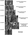
Practical examples of CAD-RADS categories using PCCT Angiography. The improved spatial resolution merged with artifact reduction enables a more comprehensive assessment of features of high-risk coronary plaque, allowing for increased diagnostic performance in quantifying luminal stenosis, low-attenuation plaques (even in the presence of heavy calcification), and spotty calcifications. The scans were conducted using a commercial whole-body Dual Source Photon-Counting CT scanner (NAEOTOM Alpha, Siemens Healthineers) with a slice thickness of 0.2/0.4 mm, a reconstruction increment of 0.1/0.2 mm, FOV 140–160 mm, and a resolution matrix of 1024 × 1024 pixels for the source axial reconstructions. Kernel filtering was set to Bv48–72 (vascular kernel medium-sharp) and Quantum Iterative Reconstruction (QIR 4) was employed with maximum intensity. The displayed image resolution is 100 microns.

References

    1. Roth G.A., Johnson C., Abajobir A., Abd-Allah F., Abera S.F., Abyu G., Ahmed M., Aksut B., Alam T., Alam K., et al. Global, Regional, and National Burden of Cardiovascular Diseases for 10 Causes, 1990 to 2015. J. Am. Coll. Cardiol. 2017;70:1–25. doi: 10.1016/j.jacc.2017.04.052. - DOI - PMC - PubMed
    1. Mathers C.D., Loncar D. Projections of global mortality and burden of disease from 2002 to 2030. PLoS Med. 2006;3:e442. doi: 10.1371/journal.pmed.0030442. - DOI - PMC - PubMed
    1. Dai H., Much A.A., Maor E., Asher E., Younis A., Xu Y., Lu Y., Liu X., Shu J., Bragazzi N.L. Global, regional, and national burden of ischaemic heart disease and its attributable risk factors, 1990-2017: Results from the Global Burden of Disease Study 2017. Eur. Heart J.-Qual. Care Clin. Outcomes. 2022;8:50–60. doi: 10.1093/ehjqcco/qcaa076. - DOI - PMC - PubMed
    1. Falk E. Pathogenesis of atherosclerosis. J. Am. Coll. Cardiol. 2006;47((Suppl. 8)):C7–C12. doi: 10.1016/j.jacc.2005.09.068. - DOI - PubMed
    1. Naghavi M., Libby P., Falk E., Casscells S.W., Litovsky S., Rumberger J., Badimon J.J., Stefanadis C., Moreno P., Pasterkamp G., et al. From Vulnerable Plaque to Vulnerable Patient. Circulation. 2003;108:1664–1672. doi: 10.1161/01.CIR.0000087480.94275.97. - DOI - PubMed

LinkOut - more resources