

A Comprehensive Evaluation of Deep Learning Models on Knee MRIs for the Diagnosis and Classification of Meniscal Tears: A Systematic Review and Meta-Analysis
- PMID: 38893617
- PMCID: PMC11172202
- DOI: 10.3390/diagnostics14111090
A Comprehensive Evaluation of Deep Learning Models on Knee MRIs for the Diagnosis and Classification of Meniscal Tears: A Systematic Review and Meta-Analysis
Abstract
Objectives: This study delves into the cutting-edge field of deep learning techniques, particularly deep convolutional neural networks (DCNNs), which have demonstrated unprecedented potential in assisting radiologists and orthopedic surgeons in precisely identifying meniscal tears. This research aims to evaluate the effectiveness of deep learning models in recognizing, localizing, describing, and categorizing meniscal tears in magnetic resonance images (MRIs).
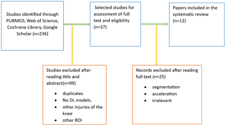
Materials and methods: This systematic review was rigorously conducted, strictly following the Preferred Reporting Items for Systematic Reviews and Meta-Analyses (PRISMA) guidelines. Extensive searches were conducted on MEDLINE (PubMed), Web of Science, Cochrane Library, and Google Scholar. All identified articles underwent a comprehensive risk of bias analysis. Predictive performance values were either extracted or calculated for quantitative analysis, including sensitivity and specificity. The meta-analysis was performed for all prediction models that identified the presence and location of meniscus tears.
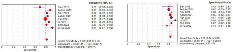
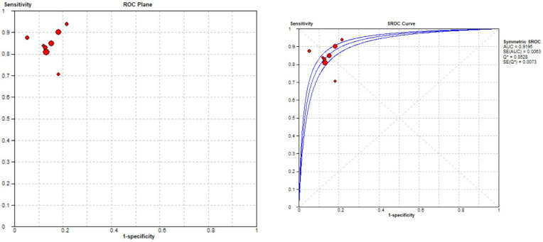
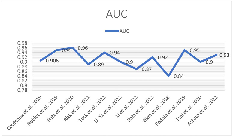
Results: This study's findings underscore that a range of deep learning models exhibit robust performance in detecting and classifying meniscal tears, in one case surpassing the expertise of musculoskeletal radiologists. Most studies in this review concentrated on identifying tears in the medial or lateral meniscus and even precisely locating tears-whether in the anterior or posterior horn-with exceptional accuracy, as demonstrated by AUC values ranging from 0.83 to 0.94.
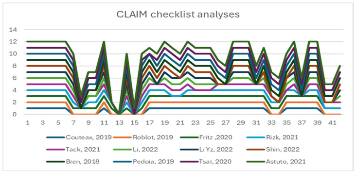
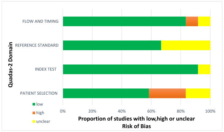
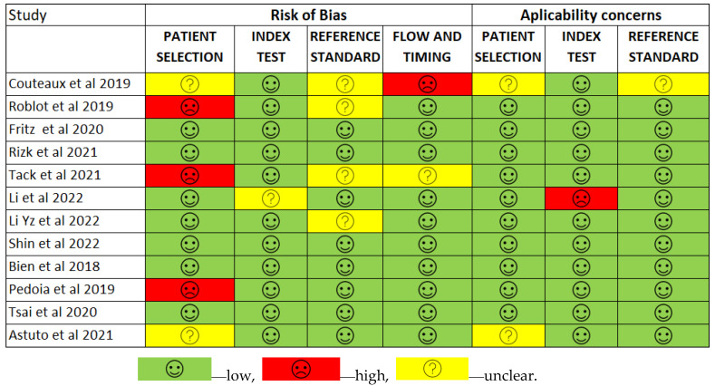
Conclusions: Based on these findings, deep learning models have showcased significant potential in analyzing knee MR images by learning intricate details within images. They offer precise outcomes across diverse tasks, including segmenting specific anatomical structures and identifying pathological regions. Contributions: This study focused exclusively on DL models for identifying and localizing meniscus tears. It presents a meta-analysis that includes eight studies for detecting the presence of a torn meniscus and a meta-analysis of three studies with low heterogeneity that localize and classify the menisci. Another novelty is the analysis of arthroscopic surgery as ground truth. The quality of the studies was assessed against the CLAIM checklist, and the risk of bias was determined using the QUADAS-2 tool.
Keywords: MRI; classification; deep learning model; diagnosis; meniscus tear.
Conflict of interest statement
The authors declare no conflicts of interest.
Figures
Similar articles
-
Artificial intelligence applied to magnetic resonance imaging reliably detects the presence, but not the location, of meniscus tears: a systematic review and meta-analysis.Eur Radiol. 2024 Sep;34(9):5954-5964. doi: 10.1007/s00330-024-10625-7. Epub 2024 Feb 22. Eur Radiol. 2024. PMID: 38386028 Free PMC article.
-
MR diagnosis of meniscal tears of the knee: importance of high signal in the meniscus that extends to the surface.AJR Am J Roentgenol. 1993 Jul;161(1):101-7. doi: 10.2214/ajr.161.1.8517286. AJR Am J Roentgenol. 1993. PMID: 8517286
-
Development of convolutional neural network model for diagnosing meniscus tear using magnetic resonance image.BMC Musculoskelet Disord. 2022 May 30;23(1):510. doi: 10.1186/s12891-022-05468-6. BMC Musculoskelet Disord. 2022. PMID: 35637451 Free PMC article.
-
Deep-learning-assisted diagnosis for knee magnetic resonance imaging: Development and retrospective validation of MRNet.PLoS Med. 2018 Nov 27;15(11):e1002699. doi: 10.1371/journal.pmed.1002699. eCollection 2018 Nov. PLoS Med. 2018. PMID: 30481176 Free PMC article.
-
Meniscal Extrusion Measurements After Posterior Medial Meniscus Root Tears: A Systematic Review and Meta-analysis.Am J Sports Med. 2023 Oct;51(12):3325-3334. doi: 10.1177/03635465221131005. Epub 2022 Dec 21. Am J Sports Med. 2023. PMID: 36541434
Cited by
-
Deep learning analysis for rheumatologic imaging: current trends, future directions, and the role of human.J Rheum Dis. 2025 Apr 1;32(2):73-88. doi: 10.4078/jrd.2024.0128. Epub 2025 Jan 20. J Rheum Dis. 2025. PMID: 40134548 Free PMC article. Review.
-
Long-Term Results for Meniscus Repair.Curr Rev Musculoskelet Med. 2025 Jul;18(7):229-245. doi: 10.1007/s12178-025-09966-7. Epub 2025 Apr 23. Curr Rev Musculoskelet Med. 2025. PMID: 40266511 Free PMC article. Review.
-
Emerging Diagnostic Approaches for Musculoskeletal Disorders: Advances in Imaging, Biomarkers, and Clinical Assessment.Diagnostics (Basel). 2025 Jun 27;15(13):1648. doi: 10.3390/diagnostics15131648. Diagnostics (Basel). 2025. PMID: 40647646 Free PMC article. Review.
-
A Geometric Deep Learning Model for Real-Time Prediction of Knee Joint Biomechanics Under Meniscal Extrusion.Ann Biomed Eng. 2025 Jul 15. doi: 10.1007/s10439-025-03798-9. Online ahead of print. Ann Biomed Eng. 2025. PMID: 40663282 Review.
-
Machine learning models for clinical and structural knee osteoarthritis prediction: Recent advancements and future directions.Osteoarthr Cartil Open. 2025 Jul 24;7(3):100654. doi: 10.1016/j.ocarto.2025.100654. eCollection 2025 Sep. Osteoarthr Cartil Open. 2025. PMID: 40799630 Free PMC article.
References
-
- Culvenor A.G., Øiestad B.E., Hart H.F., Stefanik J.J., Guermazi A., Crossley K.M. Prevalence of knee osteoarthritis features on magnetic resonance imaging in asymptomatic uninjured adults: A systematic review and meta-analysis. Br. J. Sports Med. 2019;53:1268–1278. doi: 10.1136/bjsports-2018-099257. - DOI - PMC - PubMed
Publication types
LinkOut - more resources
Full Text Sources
