Review
. 2024 May 29;14(11):1124.
doi: 10.3390/diagnostics14111124.

Ultrasound-Assisted and Ultrasound-Guided Thoracentesis: An Educational Review

Affiliations
Review

Ultrasound-Assisted and Ultrasound-Guided Thoracentesis: An Educational Review

Andrea Boccatonda et al. Diagnostics (Basel). .

Abstract

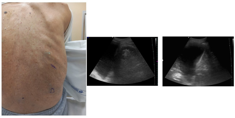
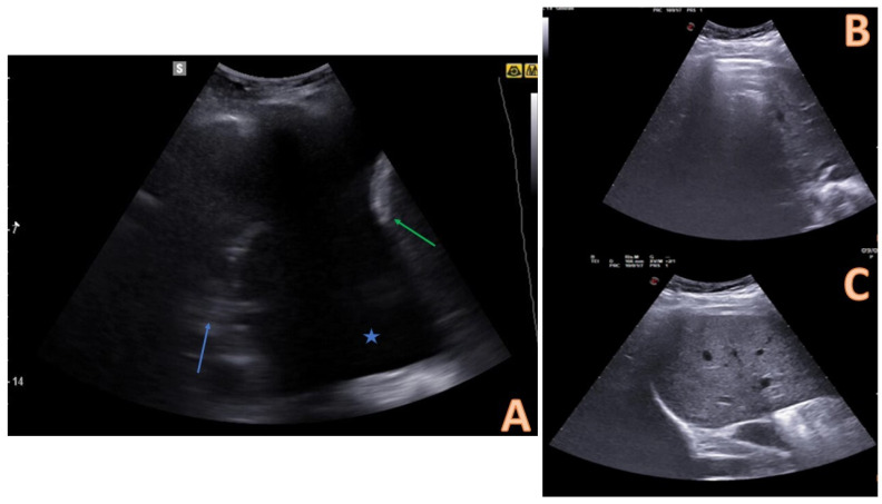
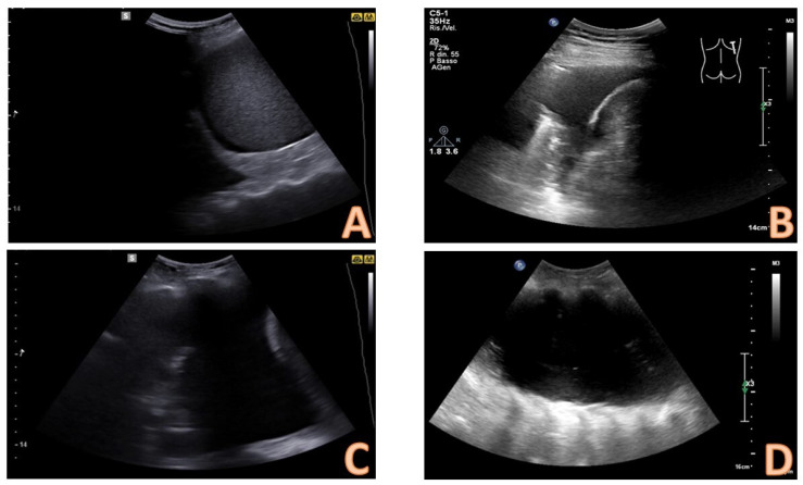
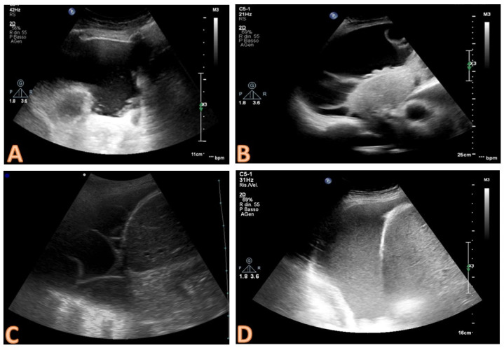
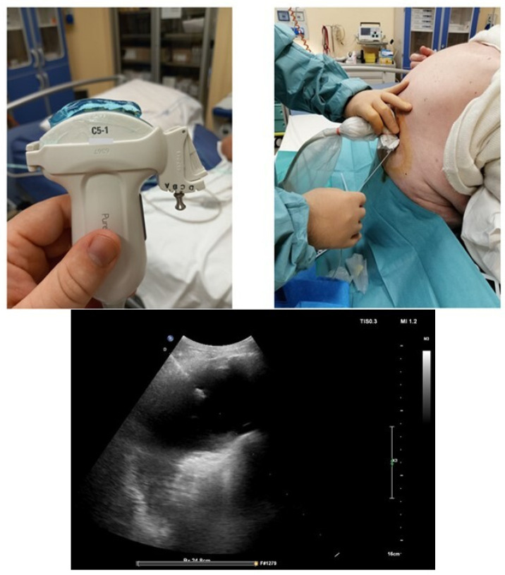
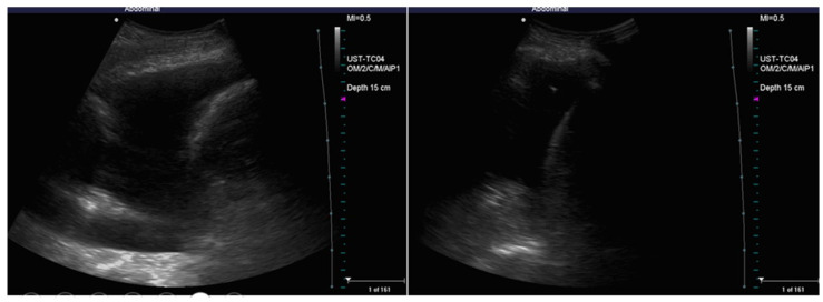
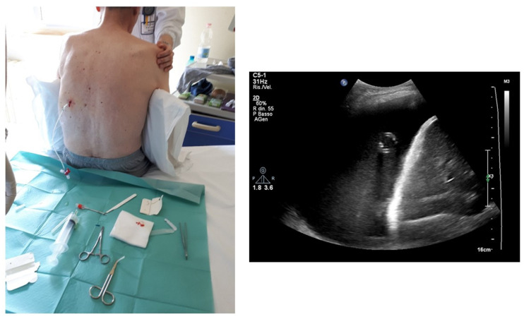
Thoracentesis is one of the most important invasive procedures in the clinical setting. Particularly, thoracentesis can be relevant in the evaluation of a new diagnosed pleural effusion, thus allowing for the collection of pleural fluid so that laboratory tests essential to establish a diagnosis can be performed. Furthermore, thoracentesis is a maneuver that can have therapeutic and palliative purposes. Historically, the procedure was performed based on a physical examination. In recent years, the role of ultrasound has been established as a valuable tool for assistance and guidance in the thoracentesis procedure. The use of ultrasound increases success rates and significantly reduces complications. The aim of this educational review is to provide a detailed and sequential examination of the procedure, focusing on the two main modalities, the ultrasound-assisted and ultrasound-guided form.

Keywords: invasive; lung; pleural effusion; procedure; thoracentesis.


Conflict of interest statement

The authors declare no conflicts of interest.

Figures

Figure 1
Figure 1
Position of the patient with posterior approach and identification of the landmarks. Ultrasound allows one to identify two landmarks: the apex of the diaphragm at the end of expiration (marked D in the image) and cranially the point (intercostal space) chosen for insertion of the needle. Ultrasound allows one to optimally identify the point where the procedure should be performed. The choice is based on the visualization of the liquid as an anechoic space. The point where the presence of anechoic fluid will be most evident should be the optimal one to perform the procedure. Often at this point the effusion takes on a “V” morphology, delimited on the sides by the diaphragm and the atelectatic lung. The apex of the “V” would represent the specific point where the needle should be inserted and directed.
Figure 2
Figure 2
Comparison between pleural effusion (A) and healthy lung on ultrasound (B,C). In (A), the pleural effusion is visible below the chest wall and ribs as an anechoic space (blue star). The effusion is delimited inferiorly by a hyperechoic formation that represents the diaphragm (green arrow), and superiorly by an echogenic structure that identifies the atelectatic lung parenchyma (blue arrow). (B,C) represent the appearance of the healthy lung base on ultrasound: an air interface with A (horizontal) lines is evident. This artefactual image covers the abdominal organ in inspirium (B) and uncovers it in expirium (B). This sign is called a “curtain sign.” When a pleural effusion is present, the curtain sign cannot be observed.
Figure 3
Figure 3
Quantitative evaluation of pleural effusion. This assessment should preferably be performed with the patient seated. It is possible to provide a semi-quantitative estimate of the effusion by counting the number of intercostal spaces occupied by the effusion through longitudinal scans starting from the diaphragm and going up cranially. In image (A), there is a minimal effusion, visible only in the costophrenic sinus; in image (B), the effusion occupies only an intercostal space and is therefore of a mild degree; in image (C). the effusion extends for 2 intercostal spaces and is therefore of moderate degree; in image (D), the entire scan is occupied by the effusion with an extension > 3 intercostal spaces and is therefore massive.
Figure 4
Figure 4
Qualitative evaluation of the effusion. The presence of echogenic material and/or septa within the effusion are suggestive of an exudate. In image (A), some echogenic and corpuscular materials are evident in the deep portion of the effusion (non-septated complex); in images (B,C), the septa are clearly evident (complex septated effusions); in image (D), the effusion appears homogeneously echogenic (the case of pleural empyema in the early phase).
Figure 5
Figure 5
Focused ultrasound evaluation of the needle insertion point. In image (A), an anomalous course of an arterial vessel in the intercostal space is evident; therefore, there is a high risk that the needle could injure the vessel, and this finding must induce the operator to vary the choice of insertion point. In image (C), a pleural plaque is evident; to insert the needle into the point where this plaque is present can both induce difficulties in inserting the needle and increasing the risk of bleeding from the plaque itself; the needle insertion point should be changed. Image (B) demonstrates how a pre-procedural ultrasound evaluation can highlight solid masses at the pleural level; this finding raises the suspicion of a malignant nature of the effusion. In image (D), there is an example of the evaluation of the distances between skin and effusion and between skin and lung, which can be done by ultrasound; this evaluation guides the choice of the needle and its insertion depth.
Figure 6
Figure 6
Example of a thoracentesis kit: the kit includes 3 needles of different caliber, a connection system with a 3-way tap, a syringe for aspiration, and a collection bag. On the left side, there is a sterile probe cover and a sterile gel pack. In the best operating setting with a mobilizable and cooperative patient, the patient sits on the edge of the bed and hugs a pillow; this movement allows the intercostal spaces to open more easily. A sterile field with a sterile drape is set up. The operator wears sterile gown and gloves. In this figure, the needle insertion site has already been identified through ultrasound evaluation, and local disinfection is carried out with povidone iodine by using sterile gauze.
Figure 7
Figure 7
Local anesthesia with lidocaine 1–2%. An ampule of lidocaine is drawn into a 10 mL syringe. In (A), the needle insertion phase is visible; the injection is performed at the chosen point and intercostal space. The non-dominant hand palpates the lower rib of the intercostal space; the dominant hand inserts the needle just above the upper edge of the lower rib; the needle is always directed downwards and never upwards to avoid damaging the intercostal artery that runs along the lower edge of the upper rib. The needle must pass the pleura and enter the pleural cavity, so that the local anesthetic can act on the richly innervated pleura; this is demonstrated by the fact that pleural fluid is aspirated into the syringe (B). At this point, boluses of 1–2 cc of anesthetic are injected at different points following a backward way to the subcutaneous tissue (C).
Figure 8
Figure 8
Sequence of images about the insertion of the needle and the execution of the thoracentesis. In image (A), the non-dominant hand palpates the lower rib of the chosen intercostal space, and the dominant hand inserts the needle above the upper edge of the rib, always directing it downwards; it should be noted that, as it is an ultrasound-assisted procedure, the probe is not held in the hand when the needle is inserted, but is available to the operator in the sterile field with a sterile probe cover. In (B,C), the leakage of the pleural fluid through the collection system is highlighted, with a second operator who sequentially aspirates and allows the fluid to flow into the collection bag. Note how the ultrasound probe is taken back into the hand and used to verify the position of the needle and the residual amount of pleural effusion.
Figure 9
Figure 9
Image highlighting the positioning of the needle inside the effusion during the procedure. The needle is represented as a hyperechoic punctiform image with some posterior artifacts within the anechoic space.
Figure 10
Figure 10
Ultrasound-guided thoracentesis. In this example, we used a guide coupled on a convex probe, which creates a pre-established needle insertion angle. An oblique intercostal scan that allows one to better highlight the pleura and the effusion is performed. The needle is displayed along its entire path, and the exact position of the tip can be observed for the entire duration of the procedure.
Figure 11
Figure 11
Sequential ultrasound check of the effusion and the position of the needle during thoracentesis. There is a progressive reduction of the anechoic space with progressive re-expansion of the atelectatic lung with the appearance of internal air bronchograms. When the tip of the needle is too close to the re-expanded lung (right figure), the procedure should be stopped.
Figure 12
Figure 12
Positioning of 10 Fr caliber pig-tail drainage. The positioning of this device allows gradual removal of the effusion, especially in massive forms, avoiding the development of barotrauma. The correct positioning of the drainage can be verified by ultrasound, viewing a track image with an oval course, corresponding to the tip of the pig-tail.

Similar articles

Cited by

References

    1. Mohammed A., Hochfeld U., Hong S., Hosseini D.K., Kim K., Omidvari K. Thoracentesis techniques: A literature review. Medicine. 2024;103:e36850. doi: 10.1097/md.0000000000036850. - DOI - PMC - PubMed
    1. Roberts M.E., Rahman N.M., Maskell N.A., Bibby A.C., Blyth K.G., Corcoran J.P., Edey A., Evison M., de Fonseka D., Hallifax R., et al. British Thoracic Society Guideline for pleural disease. Thorax. 2023;78:1143–1156. doi: 10.1136/thorax-2023-220304. - DOI - PubMed
    1. Shao R.J., Du M.J., Xie J.T. Use of lung ultrasound for the diagnosis and treatment of pleural effusion. Eur. Rev. Med. Pharmacol. Sci. 2022;26:8771–8776. doi: 10.26355/eurrev_202212_30548. - DOI - PubMed
    1. Cocco G., Boccatonda A., Rossi I., D‘Ardes D., Corvino A., Delli Pizzi A., Ucciferri C., Katia F., Jacopo V. Early detection of pleuro-pulmonary tuberculosis by bedside lung ultrasound: A case report and review of literature. Clin. Case Rep. 2022;10:e05739. doi: 10.1002/ccr3.5739. - DOI - PMC - PubMed
    1. Martinez-Zayas G., Molina S., Ost D.E. Sensitivity and complications of thoracentesis and thoracoscopy: A meta-analysis. Eur. Respir. Rev. 2022;31:220053. doi: 10.1183/16000617.0053-2022. - DOI - PMC - PubMed

LinkOut - more resources