. 2024 May 29;14(11):1126.
doi: 10.3390/diagnostics14111126.

Aging of Superficial Musculoaponeurotic System of the Face-Novel Biomarkers and Micro-CT Relevance of Facial Anti-Gravity Support

Affiliations

Aging of Superficial Musculoaponeurotic System of the Face-Novel Biomarkers and Micro-CT Relevance of Facial Anti-Gravity Support

Marius Valeriu Hînganu et al. Diagnostics (Basel). .

Abstract

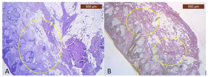
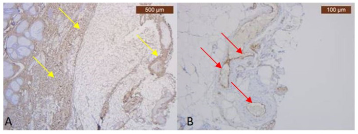
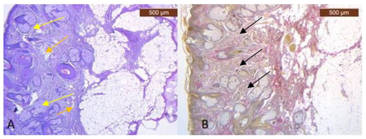
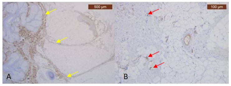
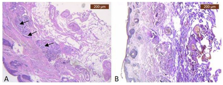
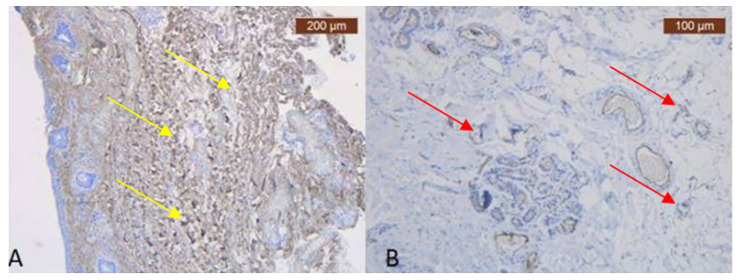
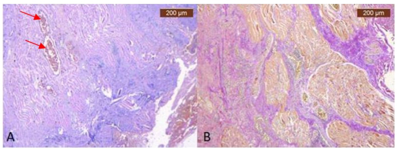
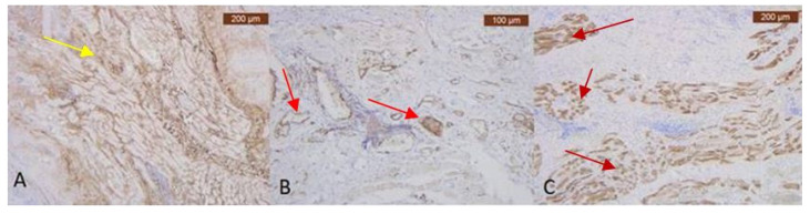
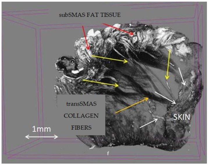
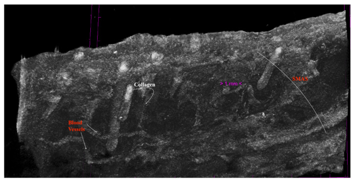
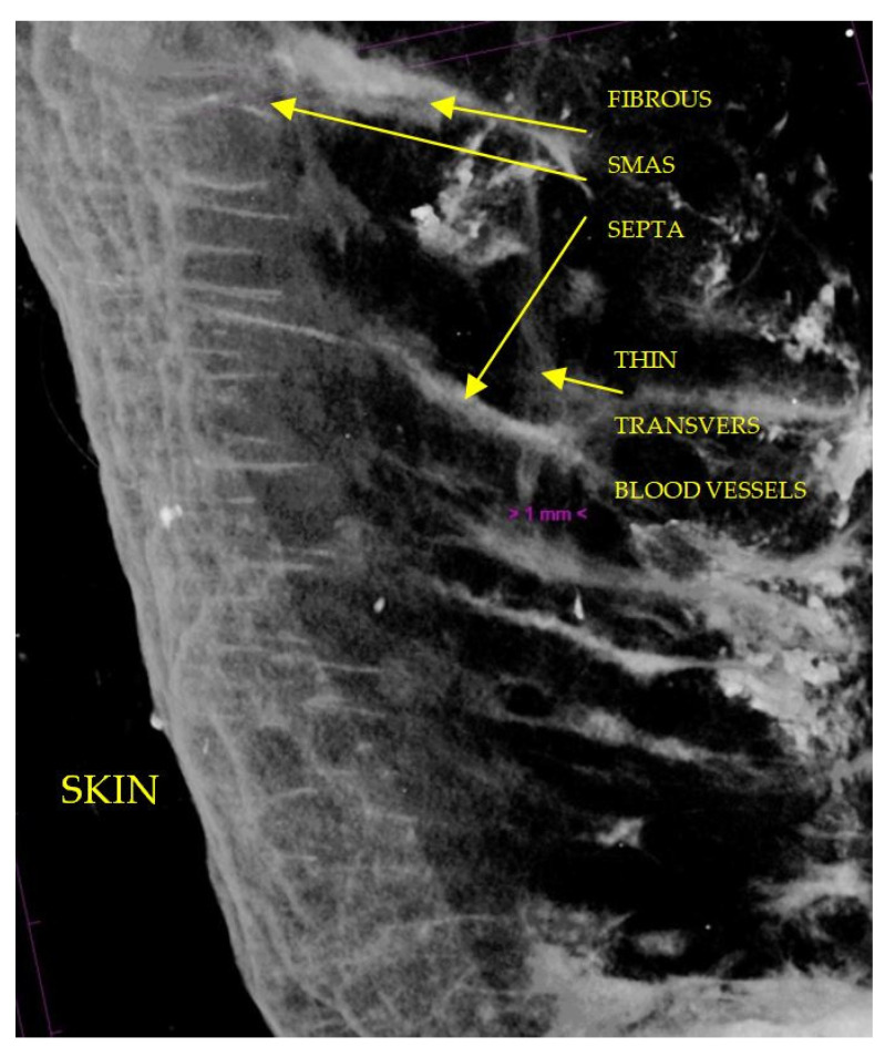
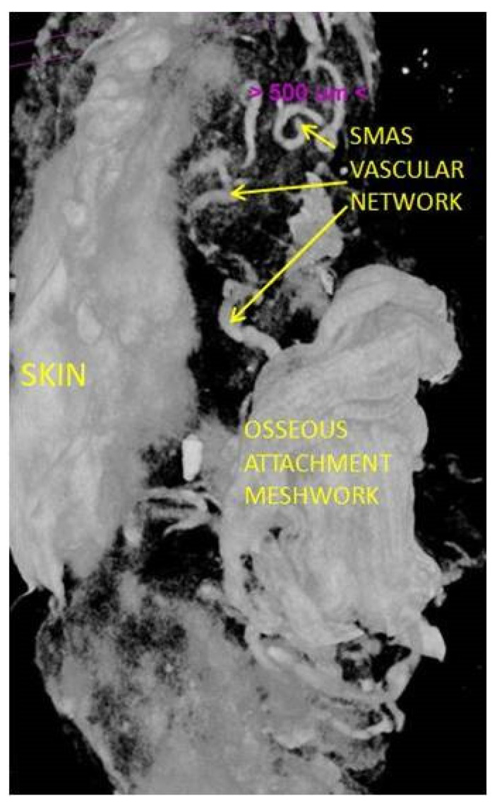
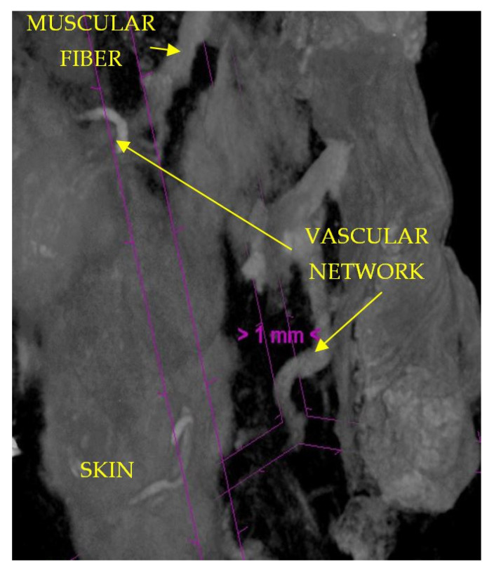
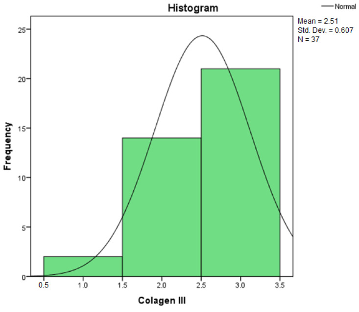
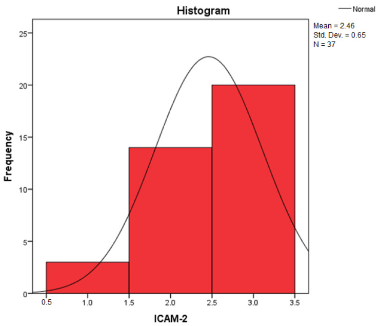
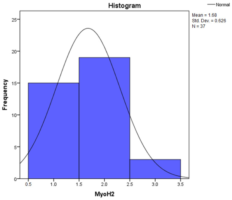
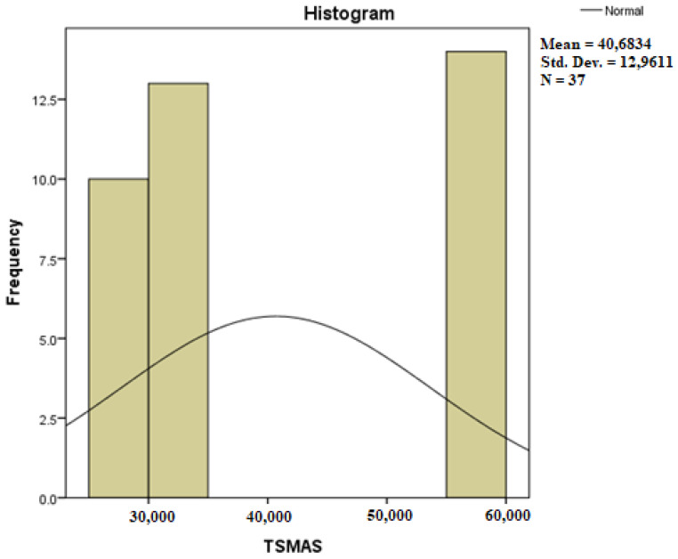
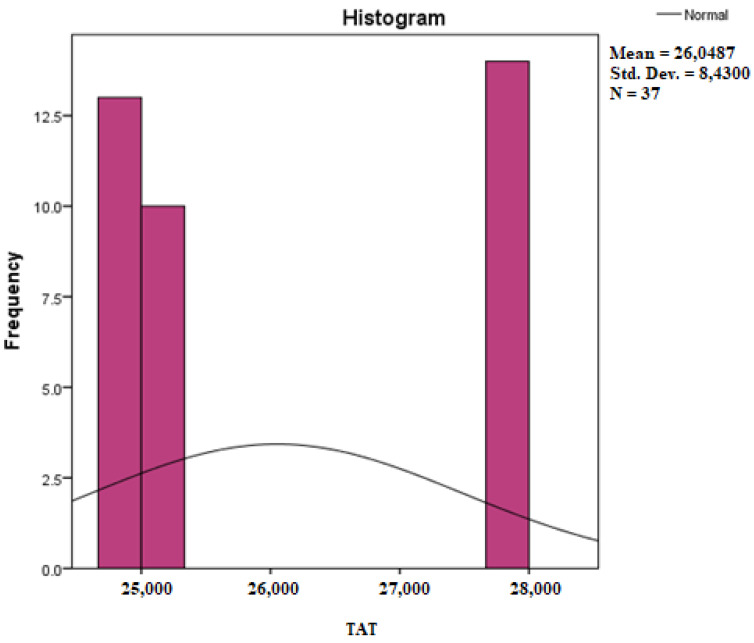
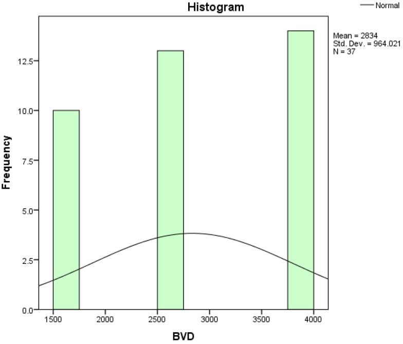
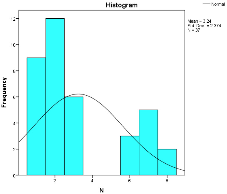
The soft superficial tissues of the face are against gravity through an intricate network of ligaments and ligamentous attachments. The aim of this investigation is to delineate the relationship between the muscular, fibrous, and vascular components of the superficial musculoaponeurotic system of the face (SMAS) at the level of its periosteal fixation areas from advanced radiological and novel biomarkers' perspectives. These areas represent key points underlying skin aging and the longevity of restorative surgery results. Methods: This study was carried out on 37 surgical specimens, excised from patients admitted for surgery. On the excised specimens, we used special immunohistochemical techniques, such as markers for collagen type III, angiogenesis, vascular endothelium (I-CAM2) and muscle fibers (MYH2). We performed a micro-CT evaluation of these 37 specimens. Results: The results of this study showed different radiologic and IHC characteristics of the means of periosteal fixation of the SMAS. Evidence of morphohistological and radiological peculiarities of the retaining ligaments highlights new data for future functional studies of these structures. Our research must be continued with larger groups of subjects and through detailed methodological studies of vascular microperfusion and could represent an important new step in biotissue engineering and the customization of surgical techniques involving the sub-SMAS layers.

Keywords: SMAS; aging biomarkers; anti-aging; facial ligaments.


Conflict of interest statement

The authors declare no conflicts of interest.

Figures

Figure 1
Figure 1
(A) Continuous musculo-adipose-fibrous network sending several extensions to the dermis (HE × 2.5); (B) Distinct fibro-adipose layer (VG × 2.5).
Figure 2
Figure 2
(A) Striking and strongly positive staining for Collagen III—yellow arrows (Anti-Collagen III × 2.5); (B) ICAM-2 mildly expressed on the blood vessels epithelium—red arrows (Anti-ICAM-2 × 10), which may be visible only in special IHC stain.
Figure 3
Figure 3
(A) Collagen (brown arrows) and adipose cells (yellow arrows) in similar proportions to the skin in deep dermis (HE × 5); (B) Superficial facial expression muscle fibers (black arrows) and adipose lobules extends to connect with the dermis of the skin (VG × 2.5).
Figure 4
Figure 4
(A) In the deep area, the collagen appeared scanty relative to that present in surrounding superficial dermal connective tissue—yellow arrows (Anti-Collagen III × 2.5); (B) ICAM is absent except in capillary and thick blood walls, which were frequently positive. Blood vessels are found only at the periphery of the lamina and are very small in size—red arrows (Anti-ICAM-2 × 10).
Figure 5
Figure 5
(A) Connective tissue layers. Nerve branches can be found in the fibrous septa in the deeper layer of the subcutaneous tissue (HE × 5)—black arrows; (B) Connective tissue layers. Fibrous septa in the deeper layer of the subcutaneous tissue (VG × 5).
Figure 6
Figure 6
(A) Type III collagen more irregularly dispersed, in the dermis—yellow arrows (Anti-Collagen III × 5); (B) Details on negative area showing staining confined to blood vessels (Anti-ICAM-2 × 10). Even in this area there are thin blood vessels within SMAS, visible on special IHC marker—red arrow.
Figure 7
Figure 7
(A) Overview of temporo-frontal area, which consist of fibromuscular sheet of delicate collagen fibrils and muscle fibers containing the vasculature—red arrows mark IHC only visible in ensheathed blood vessels (HE × 5); (B) Deep vertical and horizontal collagen fibrous septa (red) and muscular fibers (yellow) (VG × 5).
Figure 8
Figure 8
(A) Abundant perivascular and perimuscular type III collagen—yellow arrow (Anti-Collagen III × 5). Staining intensity: moderate; (B) ICAM-2 is expressed in blood vessels’ epithelium, with moderate and strong stain of endothelium—red arrows (Anti-ICAM-2 × 10). Staining intensity: weak and moderate, which highlights thin blood vessels within SMAS visible only using special IHC marker; (C) Immunohistochemically strong positive myosin reaction of muscular fibers in SMAS fibrous area—dark red arrows (Anti-MyH2 × 5). Staining intensity: moderate.
Figure 9
Figure 9
Micro-CT specimen after 7 days immersion in Lugol iodide solution. Pretragal area. 7 µm scanning; red arrows—subSMAS fat tissue; white arrows—skin; yellow arrows—blood vessels; orange arrow—collagen fibers.
Figure 10
Figure 10
SMAS fibrous collagen septa crossing to the skin together with muscular fibers; transvers and tortuous blood vessels within SMAS. Preauricular area; specimen after 14 days in Lugol Iodide solution. 7 µm scanning.
Figure 11
Figure 11
7 µm scanning of infraorbital region, rotated at 90° in order to avoid a large amount of adipose tissue and to see SMAS in detail; numerous collagen tracts of SMAS running into the skin; thin transvers blood vessels.
Figure 12
Figure 12
Well-represented vascular network and a strong collagen attaching meshwork; 14-day immersed specimen in Lugol iodide solution; 7 µm scanning.
Figure 13
Figure 13
Moderate vascular network in temporo-frontal region; moderate muscular fibers in 7-day immersed specimen in Lugol iodide solution; 7 µm scanning.
Figure 14
Figure 14
Histogram for the variable Collagen III.
Figure 15
Figure 15
Histogram for the variable ICAM-2.
Figure 16
Figure 16
Histogram for the variable MyoH2.
Figure 17
Figure 17
Histogram for the variable TSMAS.
Figure 18
Figure 18
Histogram for the variable TAT.
Figure 19
Figure 19
Histogram for the variable BVD.
Figure 20
Figure 20
Histogram for the variable N.
Figure 21
Figure 21
Histogram for the variable L.

Similar articles

Cited by

References

    1. Yadav S., Maurya P.K. Correlation Between Telomere Length and Biomarkers of Oxidative Stress in Human Aging. Rejuvenation Res. 2022;25:25–29. doi: 10.1089/rej.2021.0045. - DOI - PubMed
    1. Gu Y., Han J., Jiang C., Zhang Y. Biomarkers, oxidative stress and autophagy in skin aging. Ageing Res. Rev. 2020;59:101036. doi: 10.1016/j.arr.2020.101036. - DOI - PubMed
    1. Sahin O.S., Hanalioglu S., Metin Sanli A., Bakir A., Comert A., Baskaya M.K. Angiographic measurement of the superficial temporal artery for potential use in cerebral bypass surgery: A combined radiological and cadaveric study. Surg. Radiol. Anat. 2024;46:605–614. doi: 10.1007/s00276-024-03325-w. - DOI - PubMed
    1. Sarigul Guduk S., Cevik Cenkeri H., Derin Cicek E., Kus S. Evaluation of aging changes of the superficial fat compartments of the midface over time: A computed tomography study. J. Cosmet. Dermatol. 2022;21:1430–1435. doi: 10.1111/jocd.14292. - DOI - PubMed
    1. Mao R., Wang Y., Wang F., Zhou L., Yan S., Lu S., Shi W., Zhang Y. Identification of Four Biomarkers of Human Skin Aging by Comprehensive Single Cell Transcriptome, Transcriptome, and Proteomics. Front. Genet. 2022;13:881051. doi: 10.3389/fgene.2022.881051. - DOI - PMC - PubMed

LinkOut - more resources