

Impact of Various Non-Contrast-Enhanced MRA Techniques on Lumen Visibility in Vascular Flow Models with a Surpass Evolve Flow Diverter
- PMID: 38893675
- PMCID: PMC11171646
- DOI: 10.3390/diagnostics14111146
Impact of Various Non-Contrast-Enhanced MRA Techniques on Lumen Visibility in Vascular Flow Models with a Surpass Evolve Flow Diverter
Abstract
Background: Silent MRA has shown promising results in evaluating the stents used for intracranial aneurysm treatment. A deep learning-based denoising and deranging algorithm was recently introduced by GE HealthCare. The purpose of this study was to compare the performance of several MRA techniques regarding lumen visibility in silicone models with flow diverter stents.
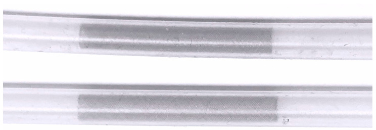
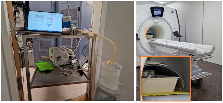
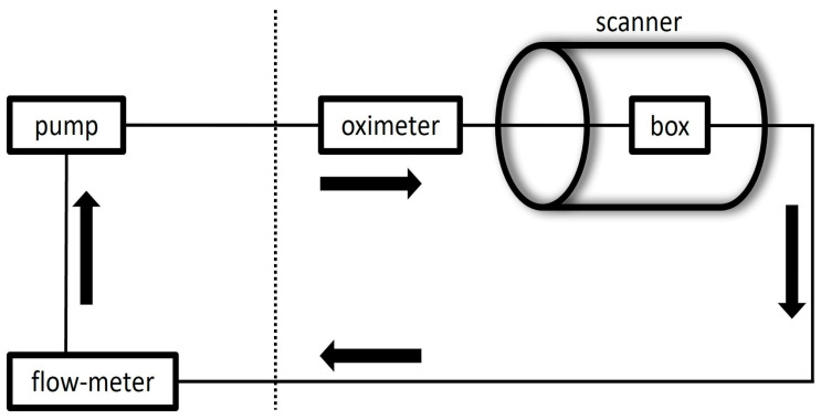
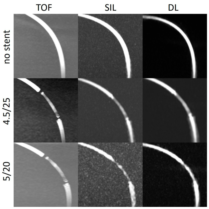
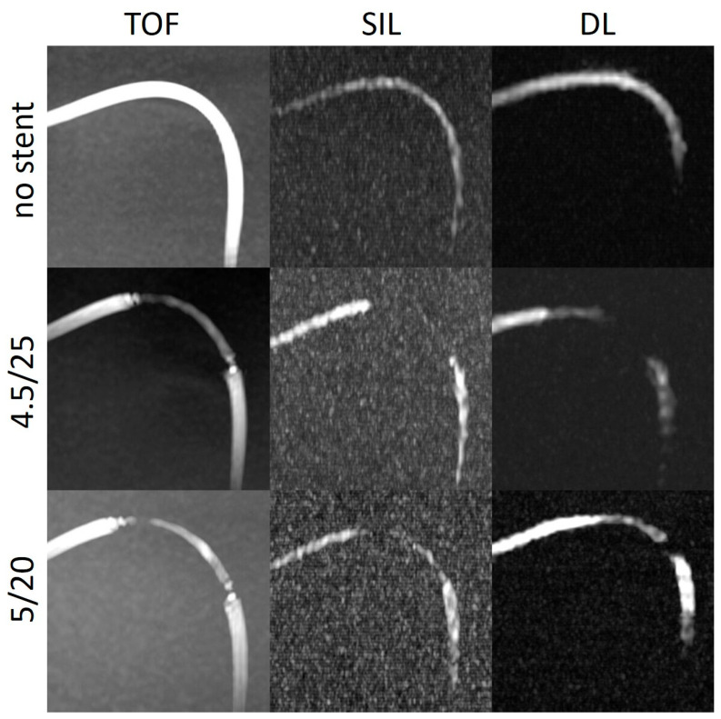
Methods: Two Surpass Evolve stents of different sizes were implanted in two silicone tubes. The tubes were placed in separate boxes in the straight position and in two different curve configurations and connected to a pulsatile pump to construct a flow loop. Using a 3.0T MRI scanner, TOF and silent MRA images were acquired, and deep learning reconstruction was applied to the silent MRA dataset. The intraluminal signal intensity in the stent (SIin-stent), in the tube outside the stent (SIvessel), and of the background (SIbg) were measured for each scan.
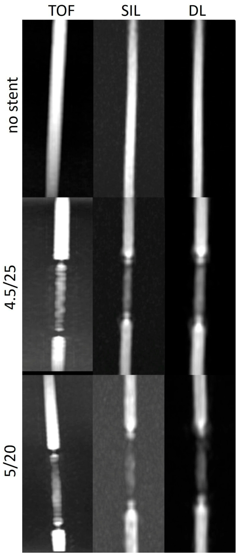
Results: The SIin-stent/SIbg and SIin-stent/SIv ratios were higher in the silent scans and DL-based reconstructions than in the TOF images. The stent tips created severe artefacts in the TOF images, which could not be observed in the silent scans.
Conclusions: Our study demonstrates that the DL reconstruction algorithm improves the quality of the silent MRA technique in evaluating the flow diverter stent patency.
Keywords: deep learning; flow diverter; intracranial aneurysm; silent MRA.
Conflict of interest statement
T.L. proctors and consults for Stryker and has in the past 3 years received service-related fees from Stryker. The other authors declare no conflicts of interest.
Figures
Similar articles
-
Assessing Blood Flow in an Intracranial Stent: A Feasibility Study of MR Angiography Using a Silent Scan after Stent-Assisted Coil Embolization for Anterior Circulation Aneurysms.AJNR Am J Neuroradiol. 2015 May;36(5):967-70. doi: 10.3174/ajnr.A4199. Epub 2014 Dec 18. AJNR Am J Neuroradiol. 2015. PMID: 25523588 Free PMC article.
-
Usefulness of Non-Contrast-Enhanced MR Angiography Using a Silent Scan for Follow-Up after Y-Configuration Stent-Assisted Coil Embolization for Basilar Tip Aneurysms.AJNR Am J Neuroradiol. 2017 Mar;38(3):577-581. doi: 10.3174/ajnr.A5033. Epub 2016 Dec 22. AJNR Am J Neuroradiol. 2017. PMID: 28007767 Free PMC article.
-
Non-Contrast-Enhanced Silent Scan MR Angiography of Intracranial Anterior Circulation Aneurysms Treated with a Low-Profile Visualized Intraluminal Support Device.AJNR Am J Neuroradiol. 2017 Aug;38(8):1610-1616. doi: 10.3174/ajnr.A5223. Epub 2017 May 18. AJNR Am J Neuroradiol. 2017. PMID: 28522664 Free PMC article.
-
Magnetic Resonance Angiography After Flow Diversion: The Use of Complementary Magnetic Resonance Angiography Techniques to Monitor Aneurysm Occlusion and Device and Arterial Branch Patency After Flow Diverter Placement.World Neurosurg. 2022 Jun;162:e147-e155. doi: 10.1016/j.wneu.2022.02.096. Epub 2022 Mar 3. World Neurosurg. 2022. PMID: 35248768
-
[Visualization of Four Intracranial Stents and In-stent Stenosis with Stenotic Models by 3D-TOF-MRA].Nihon Hoshasen Gijutsu Gakkai Zasshi. 2019;75(8):747-754. doi: 10.6009/jjrt.2019_JSRT_75.8.747. Nihon Hoshasen Gijutsu Gakkai Zasshi. 2019. PMID: 31434846 Japanese.
References
-
- Attali J., Benaissa A., Soize S., Kadziolka K., Portefaix C., Pierot L. Follow-up of intracranial aneurysms treated by flow diverter: Comparison of three-dimensional time-of-flight MR angiography (3D-TOF-MRA) and contrast-enhanced MR angiography (CE-MRA) sequences with digital subtraction angiography as the gold standard. J. Neurointerv. Surg. 2016;8:81–86. doi: 10.1136/neurintsurg-2014-011449. - DOI - PubMed
-
- Bouillot P., Brina O., Delattre B.M.A., Ouared R., Pellaton A., Yilmaz H., Machi P., Lovblad K.O., Farhat M., Pereira V.M., et al. Neurovascular stent artifacts in 3D-TOF and 3D-PCMRI: Influence of stent design on flow measurement. Magn. Reson. Med. 2019;81:560–572. doi: 10.1002/mrm.27352. - DOI - PubMed
LinkOut - more resources
Full Text Sources
