

TPMS Microarchitectures for Vertical Bone Augmentation and Osteoconduction: An In Vivo Study
- PMID: 38893806
- PMCID: PMC11173251
- DOI: 10.3390/ma17112533
TPMS Microarchitectures for Vertical Bone Augmentation and Osteoconduction: An In Vivo Study
Abstract
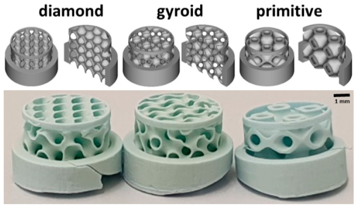
Triply periodic minimal surface microarchitectures (TPMS) were developed by mathematicians and evolved in all kingdoms of living organisms. Renowned for their lightweight yet robust attributes, TPMS structures find application in diverse fields, such as the construction of satellites, aircrafts, and electric vehicles. Moreover, these microarchitectures, despite their intricate geometric patterns, demonstrate potential for application as bone substitutes, despite the inherent gothic style of natural bone microarchitecture. Here, we produced three TPMS microarchitectures, D-diamond, G-gyroid, and P-primitive, by 3D printing from hydroxyapatite. We explored their mechanical characterization and, further, implanted them to study their bone augmentation and osteoconduction potential. In terms of strength, the D-diamond and G-gyroid performed significantly better than the P-primitive. In a calvarial defect model and a calvarial bone augmentation model, where osteoconduction is determined as the extent of bony bridging of the defect and bone augmentation as the maximal vertical bone ingrowth, the G-gyroid performed significantly better than the P-primitive. No significant difference in performance was observed between the G-gyroid and D-diamond. Since, in real life, the treatment of bone deficiencies in patients comprises elements of defect bridging and bone augmentation, ceramic scaffolds with D-diamond and G-gyroid microarchitectures appear as the best choice for a TPMS-based scaffold in bone tissue engineering.
Keywords: 3D printing; TPMS; additive manufacturing; bone substitute; ceramics; microarchitecture; osteoconduction; vertical bone augmentation.
Conflict of interest statement
The Swiss National Science Foundation (310030_197128) supported this work. The authors confirm that there are no known conflicts of interest associated with this publication and there has been no significant financial support for this work that could have influenced its outcome.
Figures


Similar articles
-
Triply Periodic Minimal Surface-Based Scaffolds for Bone Tissue Engineering: A Mechanical, In Vitro and In Vivo Study.Tissue Eng Part A. 2023 Oct;29(19-20):507-517. doi: 10.1089/ten.TEA.2023.0033. Epub 2023 Jun 19. Tissue Eng Part A. 2023. PMID: 37212290 Free PMC article.
-
Three-Dimensional Printed Hydroxyapatite Bone Substitutes Designed by a Novel Periodic Minimal Surface Algorithm Are Highly Osteoconductive.3D Print Addit Manuf. 2023 Oct 1;10(5):905-916. doi: 10.1089/3dp.2022.0134. Epub 2023 Oct 10. 3D Print Addit Manuf. 2023. PMID: 37886403 Free PMC article.
-
Hydroxyapatite 3D-printed scaffolds with Gyroid-Triply periodic minimal surface porous structure: Fabrication and an in vivo pilot study in sheep.Acta Biomater. 2023 Oct 15;170:580-595. doi: 10.1016/j.actbio.2023.08.041. Epub 2023 Sep 9. Acta Biomater. 2023. PMID: 37673232
-
Reconsidering Osteoconduction in the Era of Additive Manufacturing.Tissue Eng Part B Rev. 2019 Oct;25(5):375-386. doi: 10.1089/ten.TEB.2019.0047. Epub 2019 Sep 4. Tissue Eng Part B Rev. 2019. PMID: 30997857 Free PMC article. Review.
-
Novel 3D printed TPMS scaffolds: microstructure, characteristics and applications in bone regeneration.J Tissue Eng. 2024 Jul 26;15:20417314241263689. doi: 10.1177/20417314241263689. eCollection 2024 Jan-Dec. J Tissue Eng. 2024. PMID: 39071895 Free PMC article. Review.
Cited by
-
Compressive Behavior of Novel Additively Manufactured Ti-6Al-4V Lattice Structures: Experimental and Numerical Studies.Materials (Basel). 2024 Jul 26;17(15):3691. doi: 10.3390/ma17153691. Materials (Basel). 2024. PMID: 39124355 Free PMC article.
-
Hybrid Biomechanical Design of Dental Implants: Integrating Solid and Gyroid Triply Periodic Minimal Surface Lattice Architectures for Optimized Stress Distribution.J Funct Biomater. 2025 Feb 9;16(2):54. doi: 10.3390/jfb16020054. J Funct Biomater. 2025. PMID: 39997588 Free PMC article.
-
Strategic advances in Vat Photopolymerization for 3D printing of calcium phosphate-based bone scaffolds: A review.Bioact Mater. 2025 Jun 27;52:719-752. doi: 10.1016/j.bioactmat.2025.05.001. eCollection 2025 Oct. Bioact Mater. 2025. PMID: 40677755 Free PMC article. Review.
References
-
- Jepsen S., Schwarz F., Cordaro L., Derks J., Hämmerle C.H.F., Heitz-Mayfield L.J., Hernández-Alfaro F., Meijer H.J.A., Naenni N., Ortiz-Vigón A., et al. Regeneration of alveolar ridge defects. Consensus report of group 4 of the 15th European Workshop on Periodontology on Bone Regeneration. J. Clin. Periodontol. 2019;46((Suppl. 21)):277–286. doi: 10.1111/jcpe.13121. - DOI - PubMed
-
- Von Arx T., Cochran D.L., Hermann J.S., Schenk R.K., Buser D. Lateral ridge augmentation using different bone fillers and barrier membrane application. A histologic and histomorphometric pilot study in the canine mandible. Clin. Oral Implant. Res. 2001;12:260–269. doi: 10.1034/j.1600-0501.2001.012003260.x. - DOI - PubMed
-
- Sanz M., Dahlin C., Apatzidou D., Artzi Z., Bozic D., Calciolari E., De Bruyn H., Dommisch H., Donos N., Eickholz P., et al. Biomaterials and regenerative technologies used in bone regeneration in the craniomaxillofacial region: Consensus report of group 2 of the 15th European Workshop on Periodontology on Bone Regeneration. J. Clin. Periodontol. 2019;46((Suppl. 21)):82–91. doi: 10.1111/jcpe.13123. - DOI - PubMed
Grants and funding
LinkOut - more resources
Full Text Sources
