. 2024 Jun 6;17(11):2778.
doi: 10.3390/ma17112778.

Exploring In Vivo Pulmonary and Splenic Toxicity Profiles of Silicon Quantum Dots in Mice

Affiliations

Exploring In Vivo Pulmonary and Splenic Toxicity Profiles of Silicon Quantum Dots in Mice

Roxana-Elena Cristian et al. Materials (Basel). .

Abstract

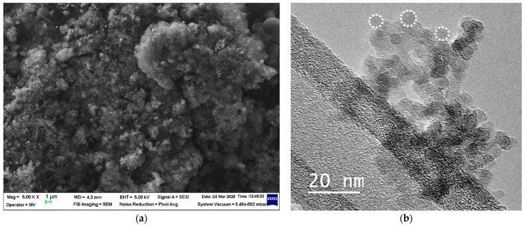
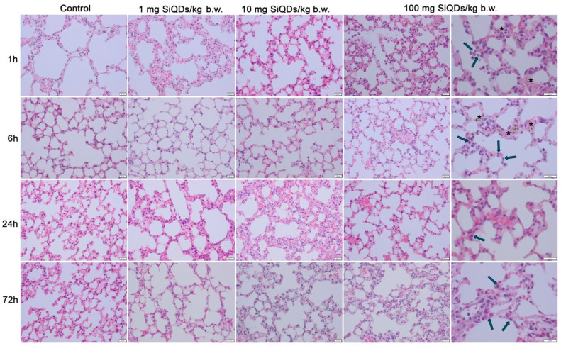
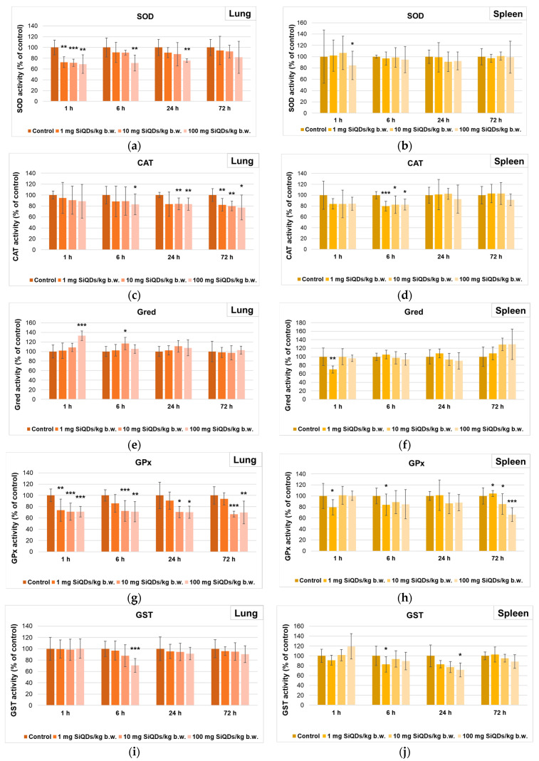
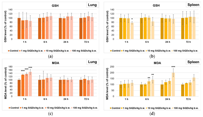
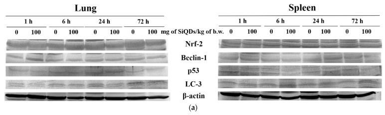
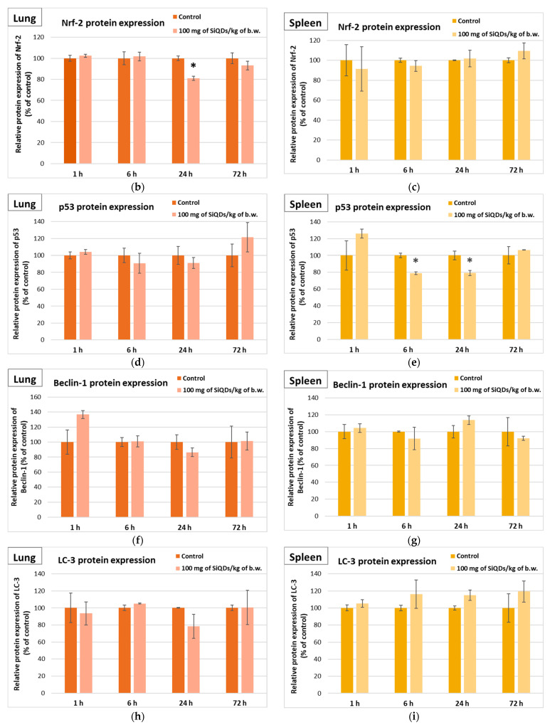
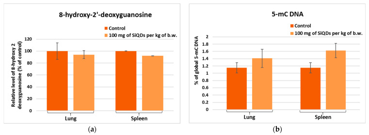
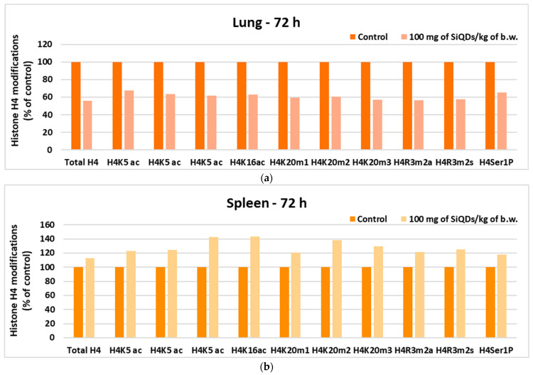
Silicon-based quantum dots (SiQDs) represent a special class of nanoparticles due to their low toxicity and easily modifiable surface properties. For this reason, they are used in applications such as bioimaging, fluorescent labeling, drug delivery, protein detection techniques, and tissue engineering despite a serious lack of information on possible in vivo effects. The present study aimed to characterize and evaluate the in vivo toxicity of SiQDs obtained by laser ablation in the lung and spleen of mice. The particles were administered in three different doses (1, 10, and 100 mg QDs/kg of body weight) by intravenous injection into the caudal vein of Swiss mice. After 1, 6, 24, and 72 h, the animals were euthanized, and the lung and spleen tissues were harvested for the evaluation of antioxidant enzyme activity, lipid peroxidation, protein expression, and epigenetic and morphological changes. The obtained results highlighted a low toxicity in pulmonary and splenic tissues for concentrations up to 10 mg SiQDs/kg body, demonstrated by biochemical and histopathological analysis. Therefore, our study brings new experimental evidence on the biocompatibility of this type of QD, suggesting the possibility of expanding research on the biomedical applications of SiQDs.

Keywords: histones; mice; oxidative stress; pulmonary and splenic toxicity; silicon quantum dots.


Conflict of interest statement

The authors declare no conflicts of interest. The funders had no role in the design of the study; in the collection, analyses, or interpretation of data; in the writing of the manuscript; or in the decision to publish the results.

Figures

Figure 1
Figure 1
The characterization of SiQDs was achieved by SEM (a) and TEM (b) analysis. The spherical shape of QDs is marked by white dot circles (b).
Figure 2
Figure 2
Histopathological evaluation of lung tissue using hematoxylin and eosin staining at intervals of 1, 6, 24, and 72 h post administration of 1 mg, 10 mg, and 100 mg of SiQDs/kg of b.w. Legend: arrows—alveolar macrophages; black stars—SiQD accumulation (brown). Scale bar (represented in the lower right corner of each image): 20 µm.
Figure 3
Figure 3
Histopathological evaluation of spleen tissue using hematoxylin and eosin staining at intervals of 1, 6, 24, and 72 h post administration of 1 mg, 10 mg, and 100 mg of SiQDs/kg of b.w. Details for 100 mg QDs/kg b.w. show the SiQD accumulation (brown dots evidenced by stars). Scale bar: 50 µm.
Figure 4
Figure 4
Specific activities of SOD (a,b), CAT (c,d), Gred (e,f), GPx (g,h), and GST (i,j) in lung (a,c,e,g,i) and spleen (b,d,f,h,j) tissues collected at 1, 6, 24, and 72 h after SiQD administration. Results are calculated as mean ± SD (n = 5) and are represented relative to the control. * p < 0.05, ** p < 0.01, and *** p < 0.001 compared to control.
Figure 5
Figure 5
Levels of GSH (a,b) and MDA (c,d) in lung (a,c) and spleen (b,d) tissues collected at 1, 6, 24, and 72 h after SiQD administration. Results are calculated as mean ± SD (n = 5) and are represented relative to the control. * p < 0.05, ** p < 0.01, and *** p < 0.001 compared to control.
Figure 6
Figure 6
Changes in proteins’ expression involved in antioxidative defense response, apoptosis, and autophagy after SiQD administration (100 mg/kg b.w.) to mice. The analysis of Nrf-2, p53 Beclin-1, and LC-3 protein expression by Western blot (a) was quantified in the lung (b,d,f,h) and spleen (c,e,g,i) tissues collected at 1, 6, 24, and 72 h after SiQD administration. Results are calculated as mean ± SD (n = 5) and are represented relative to the control. * p < 0.05 compared with the control.
Figure 6
Figure 6
Changes in proteins’ expression involved in antioxidative defense response, apoptosis, and autophagy after SiQD administration (100 mg/kg b.w.) to mice. The analysis of Nrf-2, p53 Beclin-1, and LC-3 protein expression by Western blot (a) was quantified in the lung (b,d,f,h) and spleen (c,e,g,i) tissues collected at 1, 6, 24, and 72 h after SiQD administration. Results are calculated as mean ± SD (n = 5) and are represented relative to the control. * p < 0.05 compared with the control.
Figure 7
Figure 7
The levels of 8-OHdG (a) and global DNA methylation (b) determined by ELISA technique in the murine lung and spleen samples collected at 72 h after the administration of SiQDs (100 mg QDs/kg b.w.). Results are expressed as mean ± SD (n = 5).
Figure 8
Figure 8
Changes in histone H4 in lung (a) and spleen (b) samples collected 72 h after SiQD administration (100 mg QDs/kg b.w.).

References

    1. Al-Kayiem H., Lin S., Lukmon A. Review on nanomaterials for thermal energy storage technologies. Nanosci. Nanotechnol.-Asia. 2013;3:60–71. doi: 10.2174/22113525113119990011. - DOI
    1. Soldado A., Barrio L.C., Diaz-Gonzalez M., de la Escosura-Muniz A., Costa-Fernandez J.M. Advances in quantum dots as diagnostic tools. Adv. Clin. Chem. 2022;107:1–40. doi: 10.1016/bs.acc.2021.07.001. - DOI - PubMed
    1. Ding Y., Shen S.Z., Sun H., Sun K., Liu F. Synthesis of L-glutathione-capped-ZnSe quantum dots for the sensitive and selective determination of copper ion in aqueous solutions. Sens. Actuators B Chem. 2014;203:35–43. doi: 10.1016/j.snb.2014.06.054. - DOI
    1. Nam E., Lee C., Kim S.J., Chung H.K., Chae H. Stability and dispersion improvement of quantum-dot films by hydrosilylation between quantum-dot ligands and a siloxane matrix. Opt. Express. 2019;27:20037–20046. doi: 10.1364/OE.27.020037. - DOI - PubMed
    1. Zhao Q., Rong X., Ma H., Tao G. Dithizone functionalized CdSe/CdS quantum dots as turn-on fluorescent probe for ultrasensitive detection of lead ion. J. Hazard. Mater. 2013;250:45–52. doi: 10.1016/j.jhazmat.2013.01.062. - DOI - PubMed

LinkOut - more resources