. 2024 Aug;11(31):e2400115.
doi: 10.1002/advs.202400115. Epub 2024 Jun 18.

A Novel tsRNA, m7G-3' tiRNA LysTTT, Promotes Bladder Cancer Malignancy Via Regulating ANXA2 Phosphorylation

Affiliations

A Novel tsRNA, m7G-3' tiRNA LysTTT, Promotes Bladder Cancer Malignancy Via Regulating ANXA2 Phosphorylation

Xiaoling Ying et al. Adv Sci (Weinh). 2024 Aug.

Abstract

Emerging evidence indicates that transfer RNA (tRNA)-derived small RNAs (tsRNAs), originated from tRNA with high abundance RNA modifications, play an important role in many complex physiological and pathological processes. However, the biological functions and regulatory mechanisms of modified tsRNAs in cancer remain poorly understood. Here, it is screened for and confirmed the presence of a novel m7G-modified tsRNA, m7G-3'-tiRNA LysTTT (mtiRL), in a variety of chemical carcinogenesis models by combining small RNA sequencing with an m7G small RNA-modified chip. Moreover, it is found that mtiRL, catalyzed by the tRNA m7G-modifying enzyme mettl1, promotes bladder cancer (BC) malignancy in vitro and in vivo. Mechanistically, mtiRL is found to specifically bind the oncoprotein Annexin A2 (ANXA2) to promote its Tyr24 phosphorylation by enhancing the interactions between ANXA2 and Yes proto-oncogene 1 (Yes1), leading to ANXA2 activation and increased p-ANXA2-Y24 nuclear localization in BC cells. Together, these findings define a critical role for mtiRL and suggest that targeting this novel m7G-modified tsRNA can be an efficient way for to treat BC.

Keywords: ANXA2; Bladder cancer; Yes1; m7G‐3′‐tiRNA LysTTT (mtiRL); tRNA‐derived fragments.


Conflict of interest statement

The authors declare no conflict of interest.

Figures

Figure 1
Figure 1
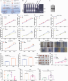
High expression of mtiRL involved in bladder cancer malignancy. A) tRNA modification was analyzed using LC/MS in multistage CdCl2 malignant transformed cells. B) Small RNA sequencing in multistage CdCl2 malignant transformed cells. C) Trend analysis of tRFs & tiRNAs. D) Model 41 in which the expression of tRFs & tiRNAs was increased upon the prolonging CdCl2 treatment time. E) Analyzed expression of m7G‐tRF&tiRNA using Arraystar Human m7G small RNA modification microarray in METTL1‐knockout T24 cells and control cells. F) m7G motif tested in Arraystar Human m7G small RNA modification microarray. G) 12 tRFs&tiRNAs containing m7G motif were significantly up‐regulated, while 94 containing m7G motif significantly down‐regulated after METTL1‐knockout. H) A Venn analysis on tRFs&tiRNAs in trend analysis model 41 and downregulated tRFs&tiRNAs. I) The abundance of m7G −3′tiRNA LysTTT‐10 (mtiRL) were detected in SV‐HUC‐1, Cd‐SV‐HUC‐1 and T24 cells by m7G immunoprecipitation‐ Stem‐loop RT PCR method. J) The Sanger sequencing of the PCR products mtiRL. K) Northern blot further confirmed the expression of mtiRL in SV‐HUC‐1 and Cd‐SV‐HUC‐1 cells. L) The results indicated that mtiRL decreased significantly in knockout METTL1 T24 cells. M) The expression of mtiRL was tested in para‐tumor (n = 7), NMIBC(n = 7) and MIBC samples(n = 7). N) The level of mtiRL was measured in 10 bladder tumor tissues from Benzopyrene‐induced multiple organ mice carcinogenesis models. O) Multi‐stage models of carcinogenesis using CdCl2 were constructed. P) Bladder cancer subtypes were determined by HE staining and the IHC expressions of Ki67and urothelial lineage markers CK5. Q) The expression of mtiRL in multi‐stage bladder tissues was tested. * p < 0.05, ** p < 0.01, *** p < 0.001, **** p < 0.0001.
Figure 2
Figure 2
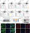
mtiRL promoted proliferation and migration of bladder cancer. A) Schematic illustration of extracting endogenous mtiRL. B) tiRNAs were separated on 12% TBE‐urea polyacrylamide gels. C) Captured endogenous mtiRL was verified by Northern blot. D–G) Cell proliferation assay showed that tiRL (without m7G modification) overexpression does not affect cell proliferation in T24 cells, METTL1‐deficient T24 cells, SV‐HUC‐1 and Cd‐SV‐HUC‐1 cells. H–K) The proliferative capacity of cells was significantly down‐regulated by small interfering RNA in T24 cells, METTL1‐deficient T24 cells, SV‐HUC‐1 and Cd‐SV‐HUC‐1 cells. L,M) Overexpression of endogenous mtiRL significantly upregulated the ability of proliferation in METTL1deficient T24 and SV‐HUC‐1 cells. N–P) Overexpression of endogenous mtiRL significantly upregulated the ability of migration in METTL1deficient T24 and SV‐HUC‐1 cells. Q–S) Both tumor volume (R) and tumor weight (S) were markedly decreased with antagomir treatment compared to NC antagomir group, whereas 3′tiRNA LysTTT overexpression did not influence tumor growth. (Each group n = 5). T) Antagomir treatment significantly reduced m7G‐3′tiRNA LysTTT expression. U) agomir treatment considerably increased 3′tiRNA LysTTT expression. * p < 0.05, ** p < 0.01, *** p < 0.001, **** p < 0.0001.
Figure 3
Figure 3
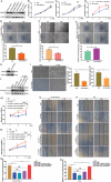
mtiRL directly binds to ANXA2. A) Schematic illustration of endogenous mtiRL pull down. B) SDS‐PAGE of pull‐down proteins. C) Venny analysis of mtiRL and tiRL pull‐down proteins. D) Protein profile of ANXA2. E) mtiRL could specifically pull down ANXA2 protein both in SV‐HUC‐1 and Cd‐SV‐HUC‐1 cells. F) RIP assays indicated that ANXA2 interacts with mtiRL. G) Domain diagram of ANXA2. H) WB detected protein expression of vectors carrying GFP‐tagged full‐length and truncated ANXA2 Flag‐tagged ANXA2. I) RNA immunoprecipitation (RIP) assays demonstrated that mtiRL interacts with the domain IV of ANXA2, but not others. All qRT‐PCR data are presented as means ±SEM. n = 3. ** p < 0.0001, **** p < 0.0001.
Figure 4
Figure 4
mtiRL increased the expression of p‐ANXA2 at Tyr24 site. A) WB results revealed that no significant difference in expression levels of ANXA2 between SV‐HUC‐1 and Cd‐SV‐HUC‐1 cells. B) ANXA2 level remained unchanged when mtiRL expression was downregulated by small interfering RNA in Cd‐SV‐HUC‐1 cells. C) WB results showed that the level of Tyr24 phosphorylation of ANXA2 (p‐ANXA2‐Y24) is up‐regulated in Cd‐SV‐HUC‐1 cells. D,E) The levels of Ser26 phosphorylation of ANXA2 (p‐ANXA2‐S26) and Tyr238 phosphorylation of ANXA2(p‐ANXA2‐Y238) were unchanged. F) The level of p‐ANXA2‐Y24 decreased with mtiRL downregulation. G,H) The expressions of p‐ANXA2‐S26 and p‐ANXA2‐Y238 remained unaltered after mtiRL downregulation. I) WB results indicated that mtiRL interfering RNA significantly downregulated p‐ANXA2‐Y24 expression in mice tumor tissues treated with antagomir. J) The results of nucleocytoplasmic separation experiment showed that mtiRL mostly distributed in nuclei. K) Nucleocytoplasmic separation assays demonstrated that ANXA2 mainly localized in cytoplasmic, while p‐ANXA2‐Y24 largely distributed in nucleus. The expression of p‐ANXA2‐Y24 reduced in Cd‐SV‐HUC‐1 cells treated with mtiRL interfering RNA. L,M) The results of immunofluorescence assays were consistent with that of nucleocytoplasmic separation assays. (Scale bar, 100 µm), * p < 0.05.
Figure 5
Figure 5
mtiRL enhances the binding between Yes1 and ANXA2. A,B) Endogenous Co‐immunoprecipitation (co‐IP) assays revealed that Yes1 bound to ANXA2. C,D) Exogenous co‐IP assays confirmed that GFP‐Yes1 interacted with Flag‐ ANXA2. E) Colocalization of Yes1 and ANXA2 was confirmed by immunofluorescence staining in Cd‐SV‐HUC‐1 cells. F–I) WB results showed that overexpression of Yes1 upregulated the level of p‐ANXA2‐Y24, whereas it did not affect the expression of total ANXA2 protein. J–M) Co‐IP experiments in Cd‐SV‐HUC‐1 cells revealed that the interaction between endogenous Yes1 and ANXA2 was significantly diminished by knockdown of mtiRL. N) In vitro kinase assay indicated that active GST‐Yes1 could phosphorylate ANXA2 at Tyr24 site and mtiRL enhance the phosphorylation of Yes1. * p < 0.05, ** p < 0.01.
Figure 6
Figure 6
ANXA2 restored proliferation and migration of mtiRL knock down cells. A). Knockout and overexpression of ANXA2 in Cd‐SV‐HUC‐1 cells were detected by WB. B,C) Cellular proliferation demonstrated that ANXA2 depletion suppressed cell proliferation. D) Over‐expression ANXA2 restored the proliferation of mtiRL‐KD cells. E,F) Scratch assays demonstrated that ANXA2 depletion suppressed cell migration ability. G) Over‐expression ANXA2 restored the migration of mtiRL‐KD cells. H–K) ANXA2 knockdown by lentivirus reduces the numbers and size of organoids from models of carcinogenesis using CdCl2. L) The wildtype (WT‐ANXA2) or MT‐ANXA2‐Y24F vectors were transfected into mtiRL‐KD cells M) Over‐expression WT‐ANXA2 but not MT‐ANXA2‐Y24F reverted the proliferation of mtiRL‐KD Cd‐SV‐HUC‐1 cells. N) Over‐expression WT‐ANXA2 but not MT‐ANXA2‐Y24F reverted the proliferation of mtiRL‐KD T24 cells. N–Q) Over‐expression WT‐ANXA2 but not MT‐ANXA2‐Y24F reverted migration phenotypes in both mtiRL‐KD Cd‐SV‐HUC‐1 cells (N,P) and mtiRL‐KD T24 cells (O,Q). * p < 0.05, ** p < 0.01, *** p < 0.001, **** p < 0.0001.
Figure 7
Figure 7
High expression level of p‐ANXA2‐Y24 is associated with a poor prognosis. A). The levels of p‐ANXA2‐Y24 in various bladder cancer cell lines and SV‐HUC‐1 cells were detected by WB. B,C) The level of p‐ANXA2‐Y24 was up‐regulated in tumor tissues compared with para‐cancer normal BC tissues. D) Kaplan–Meier survival analysis demonstrated that BC patients with high p‐ANXA2‐Y24 expression exhibited a worse prognosis than patients with low level of p‐ANXA2‐Y24. E) Summary of mtiRL promotes bladder cancer malignancy via regulating ANXA2 phosphorylation. **** p < 0.0001.

References

    1. Nombela P., Miguel‐López B., Blanco S., Mol. Cancer 2021, 20, 18. - PMC - PubMed
    1. Kimura S., Dedon P. C., Waldor M. K., Nat. Chem. Biol. 2020, 16, 964. - PMC - PubMed
    1. El Yacoubi B., Bailly M., de Crécy‐Lagard V., Annu. Rev. Gen. 2012, 46, 69. - PubMed
    1. Meng F., Zhou M., Xiao Y., Mao X., Zheng J., Lin J., Lin T., Ye Z., Cang X., Fu Y., Wang M., Guan M.‐X., Nucleic Acids Res. 2021, 49, 1075. - PMC - PubMed
    1. Liu B., Cao J., Wang X., Guo C., Liu Y., Wang T., Cell Death Dis. 2021, 13, 24. - PMC - PubMed