. 2024 Aug 12;52(14):8271-8285.
doi: 10.1093/nar/gkae519.

Distinct DNA repair mechanisms prevent formaldehyde toxicity during development, reproduction and aging

Affiliations

Distinct DNA repair mechanisms prevent formaldehyde toxicity during development, reproduction and aging

Matthias Rieckher et al. Nucleic Acids Res. .

Abstract

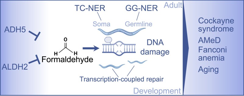
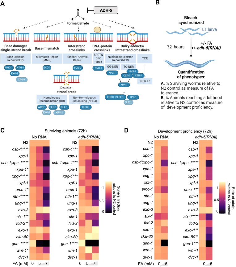
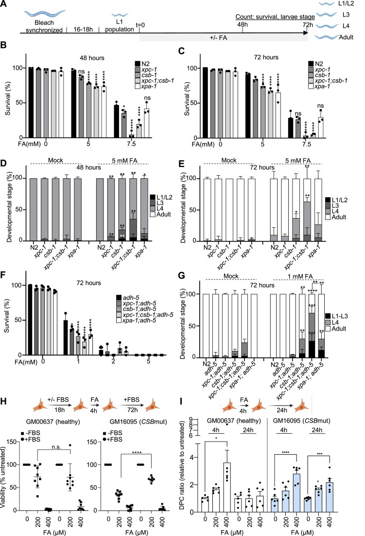
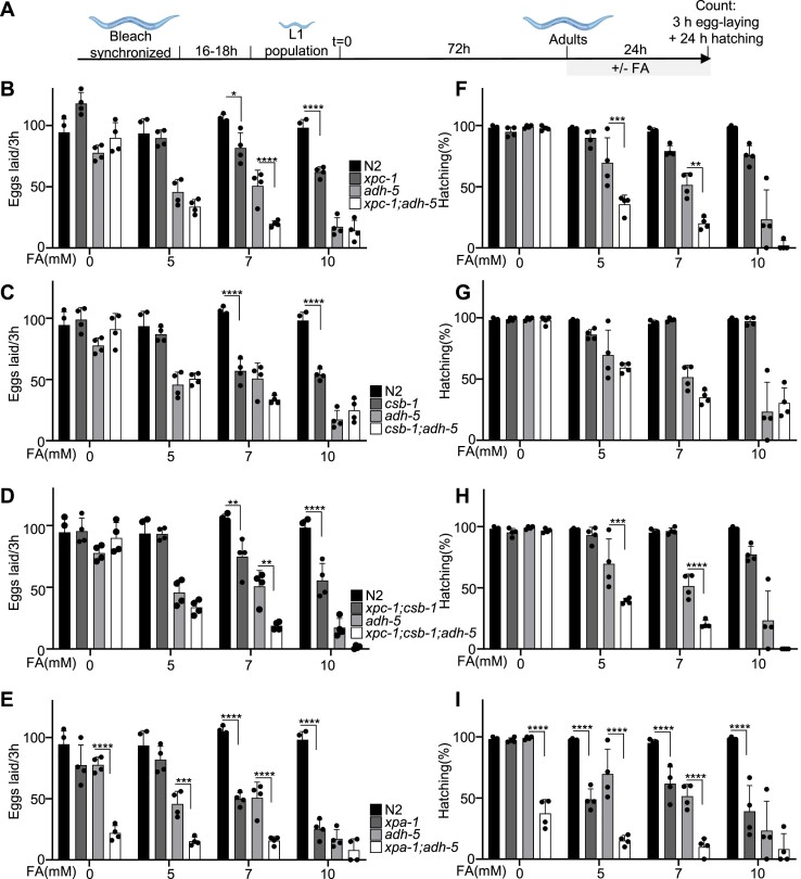
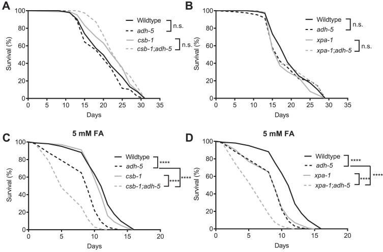
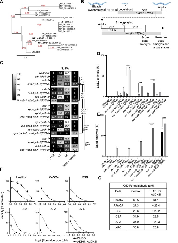
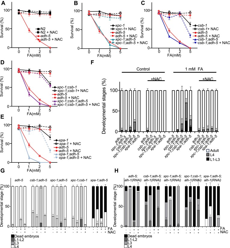
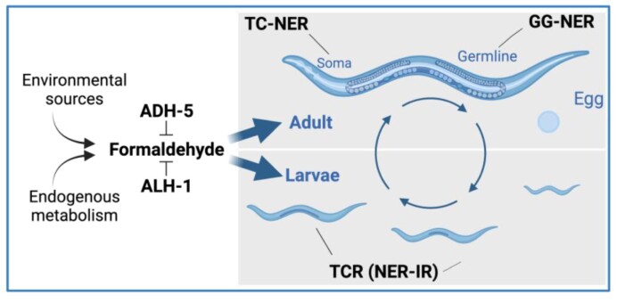
Formaldehyde (FA) is a recognized environmental and metabolic toxin implicated in cancer development and aging. Inherited mutations in the FA-detoxifying enzymes ADH5 and ALDH2 genes lead to FA overload in the severe multisystem AMeD syndrome. FA accumulation causes genome damage including DNA-protein-, inter- and intra-strand crosslinks and oxidative lesions. However, the influence of distinct DNA repair systems on organismal FA resistance remains elusive. We have here investigated the consequence of a range of DNA repair mutants in a model of endogenous FA overload generated by downregulating the orthologs of human ADH5 and ALDH2 in C. elegans. We have focused on the distinct components of nucleotide excision repair (NER) during developmental growth, reproduction and aging. Our results reveal three distinct modes of repair of FA-induced DNA damage: Transcription-coupled repair (TCR) operating NER-independently during developmental growth or through NER during adulthood, and, in concert with global-genome (GG-) NER, in the germline and early embryonic development. Additionally, we show that the Cockayne syndrome B (CSB) factor is involved in the resolution of FA-induced DNA-protein crosslinks, and that the antioxidant and FA quencher N-acetyl-l-cysteine (NAC) reverses the sensitivity of detoxification and DNA repair defects during development, suggesting a therapeutic intervention to revert FA-pathogenic consequences.


Figures

Graphical Abstract
Graphical Abstract
Figure 1.
Figure 1.
DNA repair determinants of formaldehyde tolerance in C.elegans. (A) Cartoon showing the putative lesions caused on DNA by formaldehyde (FA). In blue the factors inactivated in this work. Light blue shows some factors that might participate in multiple DNA repair pathways. (B) Scheme of the protocol applied to address FA tolerance and development in the data shown in C and D (see Materials and methods for details). (C) Heatmap summarizing survival data for C. elegans mutants in DNA repair and upon downregulation of adh-5 via RNAi. (D) Heatmap summarizing animals reaching adulthood as measure of development proficiency for C. elegans mutants in DNA repair and upon silencing of ADH-5. The heatmaps contain the mean value of three independent biological replicates for each condition. The significance was determined via a two-way ANOVA and the Dunnett's multiple comparisons test, for which counts *P < 0.05, **P < 0.01, ***P < 0.001, ****P < 0.0001. The full statistical analysis for (C) and (D) is available as supplementary tables. Cartoons were created with BioRender.com.
Figure 2.
Figure 2.
NER-deficient animals show somatic formaldehyde sensitivity, which is further enhanced by loss of ADH-5. (A) Experimental scheme (see Materials and methods for details). (B) Survival of NER mutants upon formaldehyde (FA) after 48 h and (C) after 72 h on plate. (D) Developmental timing of NER mutants upon FA after 48h on plate and (E) after 72 h on plate. (F) Survival of NER mutants in an adh-5 mutant background upon FA after 72h on plate (for N2 control the survival at 5 mM is 91.5 ± 1.9% as shown in Figure 2C). (G) Developmental timing of NER mutants in an adh-5 mutant background upon FA after 72 h on plate. Each condition was performed in three technical replicates, and the significance was determined via two-way ANOVA for survival, followed by Tukey's multiple comparison test, for which is defined *P < 0.05, **P< 0.01, ***P < 0.001, ****P < 0.0001. (H) Top, scheme depicting the experimental set up; bottom viability determined 72 h after the 4 h-FA pulse (n = 6, ****P< 0.0001 for a two-tailed t-test between the groups indicated in the figure). (I) DNA–protein crosslink (DPC) accumulation after a 4 h-FA pulse determined 24 h upon recovery for GM00637 (healthy fibroblasts, left); and for GM16095 (CSBmut fibroblasts, right) (n = 6, mean ± SEM). One-way ANOVA corrected with Dunnett test for multiple comparison (*P= 0.0109, ****P< 0.0001; *P= 0.0242, ***P= 0.0003). ANOVA tables for panels B, C, D, E and G can be found as supplementary tables. Cartoons were created with BioRender.com.
Figure 3.
Figure 3.
Fecundity is reduced in NER mutants upon formaldehyde (FA) toxicity, which is further enhanced upon loss of ADH-5. (A) Experimental scheme (see Materials and Methods for details). Panels (B–E) show the number of eggs produced by three animals within 3 h for strains carrying mutations for (B) xpc-1, (C) csb-1, (D) xpc-1;csb-1 and (E) xpa-1. Panels (F–I) display the hatching rate as counted 24 h post-egg-laying for (F) xpc-1, (G) csb-1, (H) xpc-1;csb-1 and (I) xpa-1. The graphs summarize the data of four technical replicates, and the significance was determined via the two-way ANOVA, followed by the Tukey's multiple comparison test, for which counts *P< 0.05, **P< 0.01, ***P< 0.001, ****P< 0.0001. ANOVA tables for panels B–I can be found as supplementary tables. Cartoons were created with BioRender.com
Figure 4.
Figure 4.
Loss of ADH-5 does not shorten the lifespan of animals deficient for xpa-1 and csb-1, while formaldehyde-sensitivity is increased. (A) and (B) show full lifespan experiments of wildtype and adh-5 mutants, while (A) includes csb-1 and csb-1;adh-5 mutants, and (B) includes xpa-1 and xpa-1;adh-5 deficient animals. In (C) and (D), animals were exposed to 5 mM formaldehyde (FA) from the L4 stage and lifespan was determined for (C) animals deficient in csb-1 and csb-1;adh-5, and (D) mutants for xpa-1 and xpa-1;adh-5. The significance between conditions was determined via the log-rank (Mantel-Cox) test, for which is defined ****P < 0.0001.
Figure 5.
Figure 5.
Simultaneous loss of redundant formaldehyde-detoxification pathways causes embryonic lethality and developmental delay, which is enhanced in NER-deficient animals. (A) Phylogenetic tree of C. elegans proteins showing similarity to human ALDH2. Numbers in red branch support values. (B) Experimental scheme (see Materials and methods for details). Cartoons were created with BioRender.com. (C) Heatmap combining data of development and embryonic lethality of animals exposed to alh-1(RNAi). The displayed values are the mean percentages of events counted on the plates (stages and dead embryos) after 48 h of development for three technical replicates. The significance was determined via the two-way ANOVA, followed by the Tukey's multiple comparison test, for which counts *P < 0.05, **P < 0.01, ***P < 0.001, ****P< 0.0001. A full statistical analysis between all the groups is available in the supplementary tables. (D) Percentages of L1/L2 stage animals for the various mutant backgrounds with alh-1(RNAi). (E) Percentages of dead embryos counted on plates. The significance in D and E was determined using a one-way ANOVA followed by the Tukey's multiple comparison test reporting adjusted p values for each comparison. (F) Viability plots for Epstein-Barr virus (EBV) immortalized lymphoblastic cells from patients with mutations in the genes depicted in the scheme growth. Cells were exposed to formaldehyde (FA) in presence/absence of a cocktail containing the ADH5 inhibitor N6022 (10 μM) and the ALDH2 inhibitor Disulfiram (0.1 μM) (n = 3). (G) Table showing the IC50 for FA calculated from the data in F using GraphPad.
Figure 6.
Figure 6.
NAC suppresses formaldehyde (FA)-toxicity in NER mutants and animals lacking FA-detoxification. Panels (A)–(E) show animal survival, while (F) displays the developmental stages of animals treated with formaldehyde (FA) and/or N-acetyl-L-cysteine (NAC) after 72 h. (A–F) Data were produced following the same protocol as depicted in Figure 2. A full data analysis including the two-way ANOVA followed by Tukey's multiple comparisons test for (F) is available in the supplementary tables. Panels (G) and (H) were produced according to the experimental scheme used in Figure 5 and display the larval stages and dead embryos observed on the plates 48 h after treatment with FA and/or NAC. We used two-way ANOVA followed by Tukey's multiple comparisons test to determine statistical significances and a full analysis is available in the supplementary tables.
Figure 7.
Figure 7.
Proposed model. The scheme shows the proposed distinct functions of Global Genome Nucleotide Excision Repair (GG-NER); Transcription coupled repair (TCR) independent of NER (NER-IR); and TC-NER in somatic tissues (soma), germline, and during larvae development. ADH-5 and ALH-1 are shown to prevent systemic formaldehyde (FA) toxicity throughout C. elegans life cycle. Cartoons were created with BioRender.com.

Similar articles

Cited by

References

    1. Protano C., Buomprisco G., Cammalleri V., Pocino R.N., Marotta D., Simonazzi S., Cardoni F., Petyx M., Iavicoli S., Vitali M.. The carcinogenic effects of formaldehyde occupational exposure: a systematic review. Cancers (Basel). 2022; 14:165. - PMC - PubMed
    1. Burgos-Barragan G., Wit N., Meiser J., Dingler F.A., Pietzke M., Mulderrig L., Pontel L.B., Rosado I.V., Brewer T.F., Cordell R.L.et al. .. Mammals divert endogenous genotoxic formaldehyde into one-carbon metabolism. Nature. 2017; 548:549–554. - PMC - PubMed
    1. Morellato A., Umansky C., Pontel L.. The toxic side of one-carbon metabolism and epigenetics. Redox. Biol. 2021; 40:101850. - PMC - PubMed
    1. Umansky C., Morellato A.E., Rieckher M., Scheidegger M.A., Martinefski M.R., Fernández G.A., Pak O., Kolesnikova K., Reingruber H., Bollini M.et al. .. Endogenous formaldehyde scavenges cellular glutathione resulting in redox disruption and cytotoxicity. Nat. Commun. 2022; 13:745. - PMC - PubMed
    1. Pham V.N., Bruemmer K.J., Toh J.D.W., Ge E.J., Tenney L., Ward C.C., Dingler F.A., Millington C.L., Garcia-Prieto C.A., Pulos-Holmes M.C.et al. .. Formaldehyde regulates S-adenosylmethionine biosynthesis and one-carbon metabolism. Science. 2023; 382:eabp9201. - PMC - PubMed

MeSH terms

Substances