. 2023 Oct 9;13(3):285-305.
doi: 10.1159/000533818. eCollection 2024 Jun.

Two Distinct Characteristics of Immune Microenvironment in Human Hepatocellular Carcinoma with Wnt/β-Catenin Mutations

Affiliations

Two Distinct Characteristics of Immune Microenvironment in Human Hepatocellular Carcinoma with Wnt/β-Catenin Mutations

Tomoko Aoki et al. Liver Cancer. .

Abstract

Introduction: Immunotherapy is becoming a promising approach for unresectable-hepatocellular carcinoma (HCC); the anti-tumor response is affected by the tumor microenvironment (TME). Although Wnt/β-catenin mutations are reported to cause non-inflamed phenotype, their role on TME remains controversial. We aimed to clarify the heterogeneity of immunophenotype in HCC with Wnt/β-catenin mutations.

Methods: This study includes 152 resected HCCs; mutations in the catenin beta-1, adenomatous polyposis coli, or AXIN1, or AXIN2 genes were defined as Wnt/β-catenin mutations. With hierarchical cluster analyses, TME was classified into inflamed or non-inflamed classes based on the gene expressions associated with T-cell activation. Expression profiles of molecules related to cell differentiation and biliary-stem cell markers were compared between the TME classes to investigate whether differences in tumor traits were associated with TME.

Results: Forty of 152 (26.3%) HCCs carried the Wnt/β-catenin mutations. Of these, 33 were classified as non-inflamed (33/40, 82.5%) and 7 as inflamed (7/40, 17.5%). Non-inflamed class was characterized by low number of CD3+, CD4+, and CD8+ cells on immunostaining, and high mRNA expressions of AXIN2 and GLUL, which are involved in the canonical Wnt/β-catenin signaling and hepatocyte differentiation, respectively. Non-inflamed tumors showed higher enhancement on the hepatobiliary-phase of gadolinium-ethoxybenzyl-diethylenetriamine (Gd-EOB-DTPA)-enhanced magnetic resonance imaging (MRI) compared to inflamed tumors. HCCs classified as inflamed class are revealed to have high numbers of CD3+, CD4+, and CD8+ tumor infiltrating lymphocytes on immunostaining. This class is associated with increased expression of anti-epithelial cell adhesion molecule and FOXM1 accompanied by upregulation of genes related to interferon-gamma signaling, dendritic cell migration, regulatory T cells, and myeloid-derived suppressor cell activation and recognized as low enhancement nodule on Gd-EOB-DTPA-enhanced MRI.

Conclusion: Heterogeneity of tumor traits and TME was observed in HCC with Wnt/β-catenin mutation. The potential was indicated that tumor traits and TME are determined not only by the activation of the HNF4A but also by FOXM1, both of which are downstream transcription factor of the Wnt/β-catenin signaling pathway.

Keywords: Hepatocellular carcinoma; Inflamed tumors; Non-inflamed tumors; Tumor microenvironment; Wnt/β-catenin mutation.


Conflict of interest statement

N.N. is an Editorial Board member of liver cancer. T.A., Y.K., K.S., M.M., H.C., M.T., S.H., H.I., K.U., Y.M. M.T., T.N., and K.N. have no relevant conflicts of interest to disclose. S.M. Michie Sakamoto is an Associate Editor of Liver Cancer. M.K. has received grants from Taiho Pharmaceuticals, Chugai Pharmaceuticals, Otsuka, Takeda, Sumitomo Dainippon-Sumitomo, Daiichi Sankyo, AbbVie, Astellas Pharma, and Bristol-Myers Squibb. M.K. has also received grants and personal lecture fees from Merck Sharpe and Dohme (M.S.D), Eisai, and Bayer, and is an adviser for MSD, Eisai, Bayer, Bristol-Myers Squibb, Eli Lilly, Chugai, AstraZeneca and ONO Pharmaceuticals. Masatoshi Kudo is an Editor-in-Chief of liver cancer.

Figures
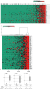
Fig. 1.
Fig. 1.
a Heatmap based on principal component analysis using the genes associated with CD8+ T cell activation. Principal component analysis was performed on gene expression for TIL activation across 152 HCCs, and a heat map was generated based on the results; cases with gene mutations such as CTNNB1 and APC were labeled. b Hierarchical cluster analysis using the genes associated with CD8+ T-cell activation. Based on the gene expression levels associated with CD8+ T cells obtained from the transcriptome analysis (see online suppl. Table 1) in 40 HCC cases with Wnt/β-catenin mutations, a cluster analysis was performed and generated a heat map with Z values. The patients were classified into two groups: those with high gene expression (inflamed class, 7 cases) and those with low gene expression (non-inflamed class, 33 cases). c Immunostaining for lymphocytes infiltrating in the tumor. Non-inflamed and inflamed classes of Wnt/β-Catenin mutated HCCs, which classified according to RNA expression levels associated with CD8+ T cells, were performed to evaluate lymphocyte infiltration into tumor. The cells stained by CD3, CD4, and CD8 were counted as cells/HPF. The inflamed class HCC showed significantly more T-cell infiltration. HCC, hepatocellular carcinoma.
Fig. 2.
Fig. 2.
Recurrence-free survival by immune class among HCCs with Wnt/β-catenin mutations. Postoperative recurrence rates were compared in 40 HCCs with Wnt/β-catenin mutations. The median RFS for the inflamed class (N = 7) was not achieved, while the median RFS for the non-inflamed class (N = 33) was 20.8 months (95% CI: 0.00–43.1). The postoperative recurrence rate was significantly lower in the inflamed class (Kaplan-Meier, Log-rank test, p = 0.018). HCC, hepatocellular carcinoma; RFS, recurrence-free survival; CI, confidence interval.
Fig. 3.
Fig. 3.
a Gene expression levels between two immune classes of HCC with and without Wnt/β-catenin mutation. Gene expression levels obtained from transcriptome analysis were compared between the two immune classes. The non-inflamed class HCC with Wnt/β-catenin mutation (N = 33) had higher expression of AXIN2 and CTNNB1, and GLUL. The inflamed class HCC with Wnt/β-catenin mutation (N = 7) had significantly lower expression of GLUL and higher expression of EMT-related genes such as SNAIL3 and VIM, TGFB1, IFN-γ, MMP9, and NOTCH3. In addition, the expression levels of chemokines such as CCL4 and CCL5, which are involved in dendritic cell migration, were significantly upregulated in the immune cold class. HCC, hepatocellular carcinoma; EMT, epithelial to mesenchymal transition; CTNNB1, catenin beta-1; GLUL, glutamate-ammonia ligase; TGFβ, transforming growth factor-β; IFN-γ, interferon gamma; MMP, matrix metalloproteinase; CCL, CC chemokine ligand; CXCL, chemokine (C-X-C motif) ligand.
Fig. 4.
Fig. 4.
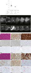
a Differences in diagnostic imaging by immunological class – signal intensity on hepatobiliary phase of Gd-EOB-DTPA-enhanced MRI – among 40 HCC patients with Wnt/β-catenin mutations, 16 patients who underwent Gd-EOB-DTPA-enhanced MRI immediately before liver resection were included in this analysis. The non-inflamed class was recognized by the presence of higher enhancement intrahepatic nodules, which were not observed in the inflamed class. b Typical Gd-EOB-DTPA-enhanced MRI study of non-inflamed and inflamed class HCCs with Wnt/β-catenin mutation; (upper) image for non-inflamed class HCC. HCC nodule was recognized as a higher enhancement on hepatobiliary phase; (lower) image for inflamed class HCC. HCC nodule was recognized as a lower enhancement on hepatobiliary phase. c–t Typical pathological evaluations of non-inflamed and inflamed class HCC with Wnt/β-catenin mutation. Non-inflamed class of HCC were as follows: (c) H&E stain, moderately differentiated HCC, (d) β-catenin stain, nuclear deposition positive, (e) GS stain, positive, (f) CK19 stain, negative, (g) EpCAM stain, negative, (h) FOXM1 stain, negative, (i) CD3 staining, (j) CD4 staining, (k) CD8 staining. Inflamed class HCC were as follows: (l) H&E stain, moderately to poorly differentiated HCC, (m) β-catenin stain, nuclear deposition positive, (n) GS stain, negative, (o) CK19 stain, 1–5% positive, (p) EpCAM stain, positive, (q) FOXM1 stain, positive, (r) CD3 staining, (s) CD4 staining, (t) CD8 staining. The red arrows indicate positive findings. H&E, hematoxylin and eosin; HCC, hepatocellular carcinoma; GS, glutamine synthetase; CK19, cytokeratin 19; EpCAM, anti-epithelial cell adhesion molecule; FOX, forkhead box; Gd-EOB-DTPA, gadolinium-ethoxybenzyl-diethylenetriamine; MRI, magnetic resonance imaging; RER, relative enhancement ratio; CI, confidence interval.
Fig. 5.
Fig. 5.
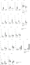
Tumor immune microenvironment and immunohistochemistry results for each immune class. In HCC with Wnt/β-catenin mutations, nuclear deposition of β-catenin could be confirmed by IHC in some patients, but not in others. In the non-inflamed class, there were significantly more HCC nodules with clear GS staining (63.6% vs. 14.3%, p = 0.047), and most cases were negative for biliary stem cell markers such as EpCAM and CK19. Conversely, the inflamed class showed a high degree of CD8+ T-cell infiltration and expression of suppressor molecules such as PD-1, LAG-3, and TIM-3. GS was rarely stained and FOXM1-positive HCC cells were more frequently observed (see Table 2). As shown in online supplementary Figure 6, cluster analysis was performed based on gene expression levels associated with NK cells, dendritic cells, immune escape, and immunosuppressor cells (see online suppl. Table 1), and the results of these axes were described as one panel an immunological TME. In each axis, two groups were reflected in the activation and non-activation groups (three groups only for IFN-γ signaling). The non-inflamed class of Wnt/β-catenin mutated HCCs was also a non-activated group in the other axes related to tumor immunity. HCC, hepatocellular carcinoma; IHC, immunohistochemistry; FOX, forkhead box; EpCAM, anti-epithelial cell adhesion molecule; CK19, cytokeratin 19; PD-1, programmed cell death protein 1; LAG-3, lymphocyte-activation gene-3; TIM-3, T-cell immunoglobulin mucin-3; GS, glutamine synthetase; HIF, hypoxia inducible factor; NK, natural killer; TME, tumor microenvironment; IFN-γ, interferon gamma.

References

    1. Takayama T, Yamazaki S, Matsuyama Y, Midorikawa Y, Shiina S; Liver Cancer Study Group of Japan, et al. . Prognostic grade for resecting hepatocellular carcinoma: multicentre retrospective study. Br J Surg. 2021 Apr 30;108(4):412–8. - PubMed
    1. Finn RS, Qin S, Ikeda M, Galle PR, Ducreux M, Kim TY, et al. . Atezolizumab plus bevacizumab in unresectable hepatocellular carcinoma. N Engl J Med Overseas Ed. 2020 May 14;382(20):1894–905. - PubMed
    1. Pinyol R, Sia D, Llovet JM. Immune exclusion-wnt/CTNNB1 class predicts resistance to immunotherapies in HCC. Clin Cancer Res. 2019 Apr 1;25(7):2021–3. - PMC - PubMed
    1. Harding JJ, Nandakumar S, Armenia J, Khalil DN, Albano M, Ly M, et al. . Prospective genotyping of hepatocellular carcinoma: clinical implications of next-generation sequencing for matching patients to targeted and immune therapies. Clin Cancer Res. 2019 Apr 1;25(7):2116–26. - PMC - PubMed
    1. Ruiz de Galarreta M, Bresnahan E, Molina-Sanchez P, Lindblad KE, Maier B, Sia D, et al. . β-Catenin activation promotes immune escape and resistance to anti-PD-1 therapy in hepatocellular carcinoma. Cancer Discov. 2019 Aug;9(8):1124–41. - PMC - PubMed