

The extracellular serine protease from Staphylococcus epidermidis elicits a type 2-biased immune response in atopic dermatitis patients
- PMID: 38895118
- PMCID: PMC11183529
- DOI: 10.3389/fimmu.2024.1352704
The extracellular serine protease from Staphylococcus epidermidis elicits a type 2-biased immune response in atopic dermatitis patients
Abstract
Background: Atopic dermatitis (AD) is a chronic, relapsing inflammatory skin disease with skin barrier defects and a misdirected type 2 immune response against harmless antigens. The skin microbiome in AD is characterized by a reduction in microbial diversity with a dominance of staphylococci, including Staphylococcus epidermidis (S. epidermidis).
Objective: To assess whether S. epidermidis antigens play a role in AD, we screened for candidate allergens and studied the T cell and humoral immune response against the extracellular serine protease (Esp).
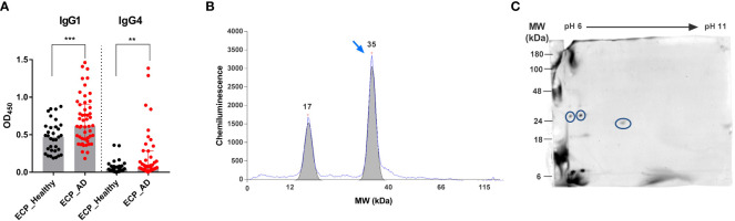
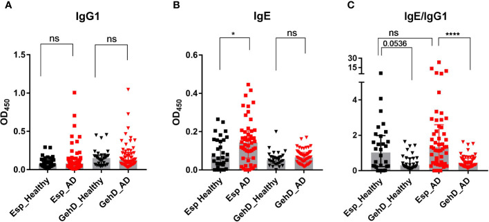
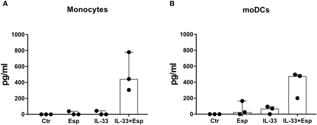
Methods: To identify candidate allergens, we analyzed the binding of human serum IgG4, as a surrogate of IgE, to S. epidermidis extracellular proteins using 2-dimensional immunoblotting and mass spectrometry. We then measured serum IgE and IgG1 binding to recombinant Esp by ELISA in healthy and AD individuals. We also stimulated T cells from AD patients and control subjects with Esp and measured the secreted cytokines. Finally, we analyzed the proteolytic activity of Esp against IL-33 and determined the cleavage sites by mass spectrometry.
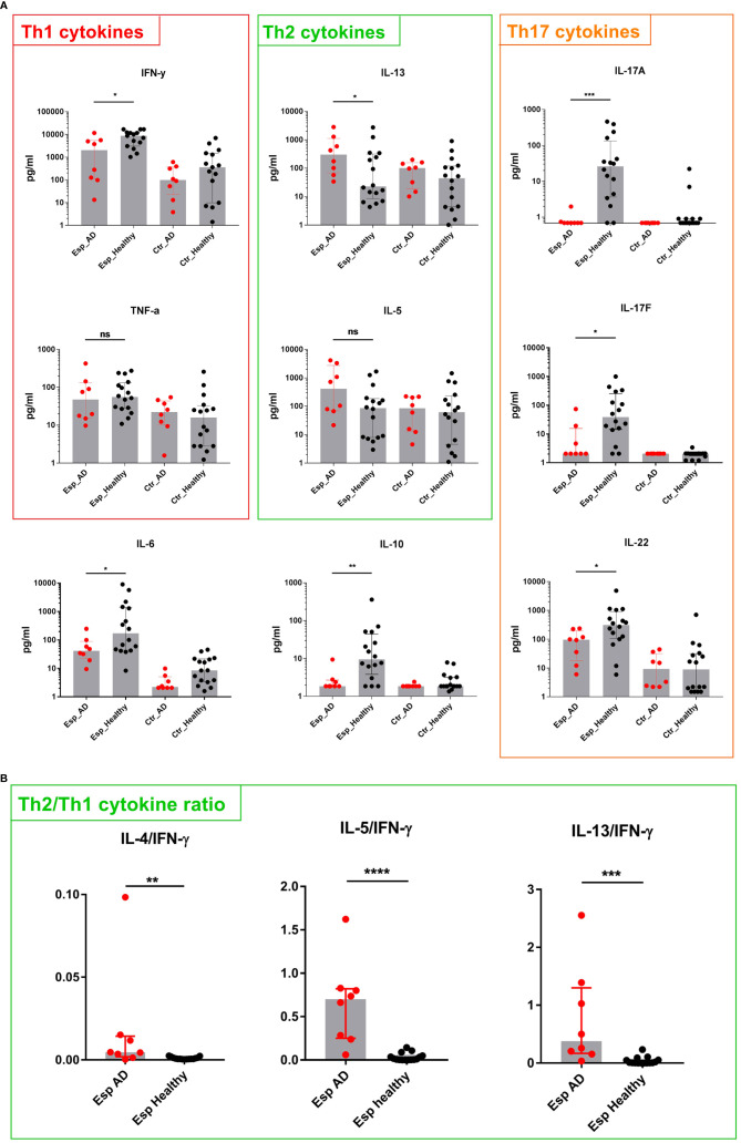
Results: We identified Esp as the dominant candidate allergen of S. epidermidis. Esp-specific IgE was present in human serum; AD patients had higher concentrations than controls. T cells reacting to Esp were detectable in both AD patients and healthy controls. The T cell response in healthy adults was characterized by IL-17, IL-22, IFN-γ, and IL-10, whereas the AD patients' T cells lacked IL-17 production and released only low amounts of IL-22, IFN-γ, and IL-10. In contrast, Th2 cytokine release was higher in T cells from AD patients than from healthy controls. Mature Esp cleaved and activated the alarmin IL-33.
Conclusion: The extracellular serine protease Esp of S. epidermidis can activate IL-33. As an antigen, Esp elicits a type 2-biased antibody and T cell response in AD patients. This suggests that S. epidermidis can aggravate AD through the allergenic properties of Esp.
Keywords: Esp; IgE; Staphylococcus epidermidis; Th2 cells; allergy; atopic dermatitis; protease.
Copyright © 2024 Abdurrahman, Pospich, Steil, Gesell Salazar, Izquierdo González, Normann, Mrochen, Scharf, Völker, Werfel, Bröker, Roesner and Gómez-Gascón.
Conflict of interest statement
The authors declare that the research was conducted in the absence of any commercial or financial relationships that could be construed as a potential conflict of interest.
Figures

Similar articles
-
Staphylococcus aureus from atopic dermatitis skin alters cytokine production triggered by monocyte-derived Langerhans cell.J Dermatol Sci. 2017 Dec;88(3):271-279. doi: 10.1016/j.jdermsci.2017.08.001. Epub 2017 Aug 5. J Dermatol Sci. 2017. PMID: 28822698
-
Staphylococcal serine protease-like proteins are pacemakers of allergic airway reactions to Staphylococcus aureus.J Allergy Clin Immunol. 2017 Feb;139(2):492-500.e8. doi: 10.1016/j.jaci.2016.03.045. Epub 2016 May 10. J Allergy Clin Immunol. 2017. PMID: 27315768
-
Candida albicans mannan- and protein-induced humoral, cellular and cytokine responses in atopic dermatitis patients.Clin Exp Allergy. 1999 Jun;29(6):824-31. doi: 10.1046/j.1365-2222.1999.00555.x. Clin Exp Allergy. 1999. PMID: 10336600
-
The adaptive immune system in atopic dermatitis and implications on therapy.Expert Rev Clin Immunol. 2016 Jul;12(7):787-96. doi: 10.1586/1744666X.2016.1165093. Epub 2016 Mar 28. Expert Rev Clin Immunol. 2016. PMID: 26967382 Review.
-
Atopic dermatitis and cytokines: recent patents in immunoregulatory and therapeutic implications of cytokines in atopic dermatitis--part I: cytokines in atopic dermatitis.Recent Pat Inflamm Allergy Drug Discov. 2012 Sep;6(3):222-47. doi: 10.2174/187221312802652820. Recent Pat Inflamm Allergy Drug Discov. 2012. PMID: 22827753 Review.
Cited by
-
Unraveling the gut-skin axis in atopic dermatitis: exploiting insights for therapeutic strategies.Gut Microbes. 2024 Jan-Dec;16(1):2430420. doi: 10.1080/19490976.2024.2430420. Epub 2024 Nov 27. Gut Microbes. 2024. PMID: 39601281 Free PMC article. Review.
-
Staphylococcus Strains in Atopic Dermatitis in Children: Toxins Production and Resistance Properties.Life (Basel). 2025 Jul 17;15(7):1120. doi: 10.3390/life15071120. Life (Basel). 2025. PMID: 40724622 Free PMC article.
References
MeSH terms
Substances
LinkOut - more resources
Full Text Sources
Research Materials