

Transcranial focused ultrasound to the posterior cingulate cortex modulates default mode network and subjective experience: an fMRI pilot study
- PMID: 38895168
- PMCID: PMC11184145
- DOI: 10.3389/fnhum.2024.1392199
Transcranial focused ultrasound to the posterior cingulate cortex modulates default mode network and subjective experience: an fMRI pilot study
Abstract
Background: Transcranial focused ultrasound (TFUS) is an emerging neuromodulation tool for temporarily altering brain activity and probing network functioning. The effects of TFUS on the default mode network (DMN) are unknown.
Objective: The study examined the effects of transcranial focused ultrasound (TFUS) on the functional connectivity of the default mode network (DMN), specifically by targeting the posterior cingulate cortex (PCC). Additionally, we investigated the subjective effects of TFUS on mood, mindfulness, and self-related processing.
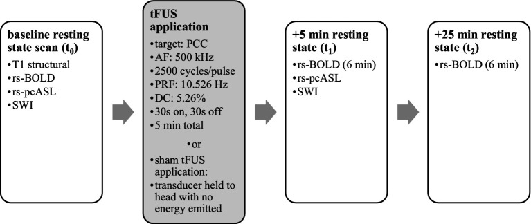
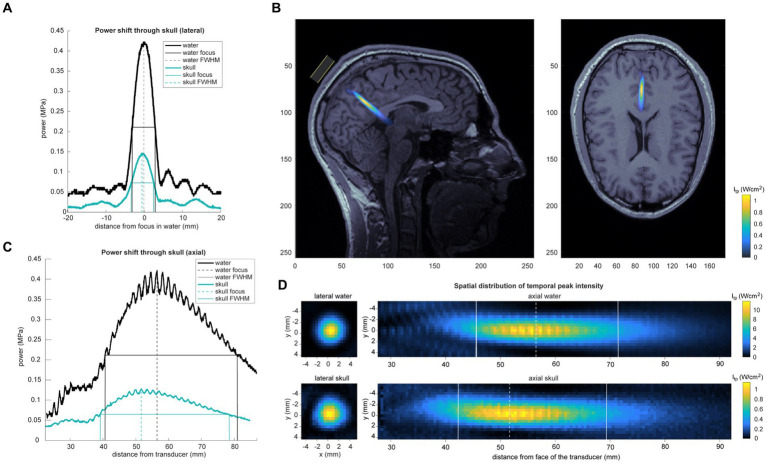
Methods: The study employed a randomized, single-blind design involving 30 healthy subjects. Participants were randomly assigned to either the active TFUS group or the sham TFUS group. Resting-state functional magnetic resonance imaging (rs-fMRI) scans were conducted before and after the TFUS application. To measure subjective effects, the Toronto Mindfulness Scale, the Visual Analog Mood Scale, and the Amsterdam Resting State Questionnaire were administered at baseline and 30 min after sonication. The Self Scale and an unstructured interview were also administered 30 min after sonication.
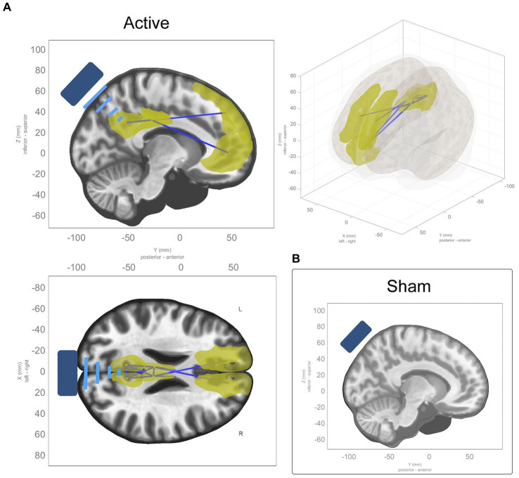
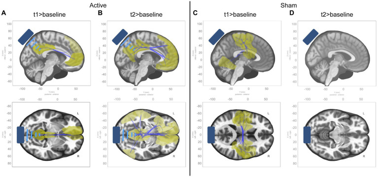
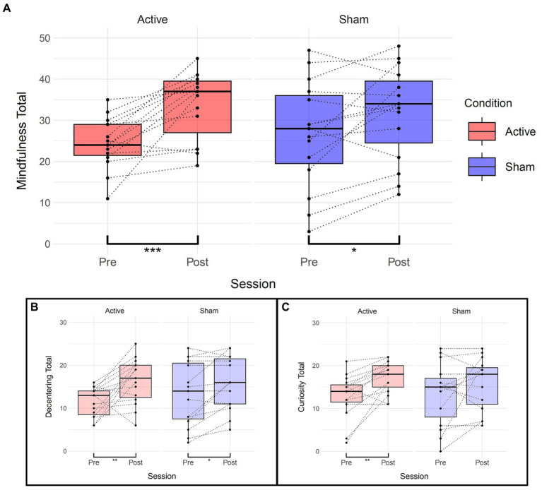
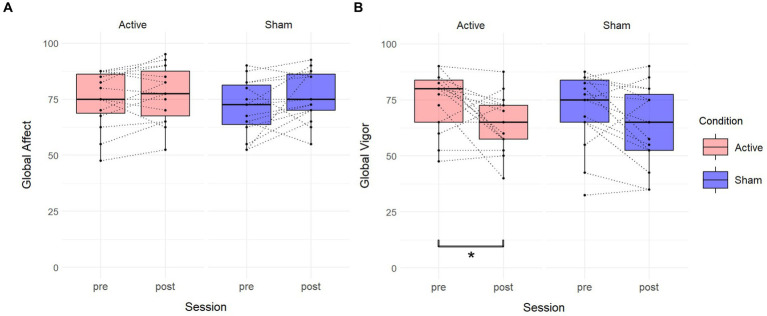
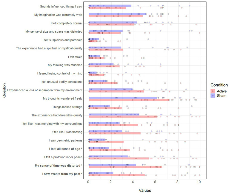
Results: The active TFUS group exhibited significant reductions in functional connectivity along the midline of the DMN, while the sham TFUS group showed no changes. The active TFUS group demonstrated increased state mindfulness, reduced Global Vigor, and temporary alterations in the sense of ego, sense of time, and recollection of memories. The sham TFUS group showed an increase in state mindfulness, too, with no other subjective effects.
Conclusions: TFUS targeted at the PCC can alter DMN connectivity and cause changes in subjective experience. These findings support the potential of TFUS to serve both as a research tool and as a potential therapeutic intervention.
Keywords: default mode network; fMRI; mindfulness; neuromodulation; non-invasive brain stimulation; transcranial focused ultrasound.
Copyright © 2024 Lord, Sanguinetti, Ruiz, Miskovic, Segre, Young, Fini and Allen.
Conflict of interest statement
JLS was paid a salary and was a shareholder in Sanmai Technologies, PBC. LR was paid a salary by Sanmai Technologies, PBC. JS and VM were paid a salary by X Moonshot Factory. The remaining authors declare that the research was conducted in the absence of any commercial or financial relationships that could be construed as a potential conflict of interest.
Figures
Similar articles
-
Alterations in large-scale resting-state network nodes following transcranial focused ultrasound of deep brain structures.Front Hum Neurosci. 2024 Dec 4;18:1486770. doi: 10.3389/fnhum.2024.1486770. eCollection 2024. Front Hum Neurosci. 2024. PMID: 39698148 Free PMC article.
-
Non-invasive suppression of the human nucleus accumbens (NAc) with transcranial focused ultrasound (tFUS) modulates the reward network: a pilot study.Front Hum Neurosci. 2024 Apr 2;18:1359396. doi: 10.3389/fnhum.2024.1359396. eCollection 2024. Front Hum Neurosci. 2024. PMID: 38628972 Free PMC article.
-
Transcranial focused ultrasound of the amygdala modulates fear network activation and connectivity.Brain Stimul. 2024 Mar-Apr;17(2):312-320. doi: 10.1016/j.brs.2024.03.004. Epub 2024 Mar 4. Brain Stimul. 2024. PMID: 38447773 Clinical Trial.
-
Resting-state fMRI functional connectivity and mindfulness in clinical and non-clinical contexts: A review and synthesis.Neurosci Biobehav Rev. 2022 Apr;135:104583. doi: 10.1016/j.neubiorev.2022.104583. Epub 2022 Feb 22. Neurosci Biobehav Rev. 2022. PMID: 35202647 Free PMC article. Review.
-
Mindfulness in the focus of the neurosciences - The contribution of neuroimaging to the understanding of mindfulness.Front Behav Neurosci. 2022 Oct 17;16:928522. doi: 10.3389/fnbeh.2022.928522. eCollection 2022. Front Behav Neurosci. 2022. PMID: 36325155 Free PMC article.
Cited by
-
Novel NIBS in psychiatry: Unveiling TUS and TI for research and treatment.Brain Neurosci Adv. 2025 Mar 14;9:23982128251322241. doi: 10.1177/23982128251322241. eCollection 2025 Jan-Dec. Brain Neurosci Adv. 2025. PMID: 40092509 Free PMC article. Review.
-
Magnitude preparation-based MR-acoustic radiation force imaging.Magn Reson Med. 2025 Oct;94(4):1445-1457. doi: 10.1002/mrm.30562. Epub 2025 May 30. Magn Reson Med. 2025. PMID: 40443184
-
Mindfulness-based neurofeedback: A systematic review of EEG and fMRI studies.Imaging Neurosci (Camb). 2024 Dec 20;2:imag-2-00396. doi: 10.1162/imag_a_00396. eCollection 2024. Imaging Neurosci (Camb). 2024. PMID: 40800428 Free PMC article.
-
Neuromodulation with Ultrasound: Hypotheses on the Directionality of Effects and Community Resource.medRxiv [Preprint]. 2025 Feb 21:2024.06.14.24308829. doi: 10.1101/2024.06.14.24308829. medRxiv. 2025. PMID: 38947047 Free PMC article. Preprint.
-
Transcranial focused ultrasound targeting the default mode network for the treatment of depression.Front Psychiatry. 2025 Apr 4;16:1451828. doi: 10.3389/fpsyt.2025.1451828. eCollection 2025. Front Psychiatry. 2025. PMID: 40256163 Free PMC article.
References
LinkOut - more resources
Full Text Sources