[Preprint]. 2024 Jun 9:2024.06.07.597791.
doi: 10.1101/2024.06.07.597791.

Diminished Immune Cell Adhesion in Hypoimmune ICAM-1 Knockout Pluripotent Stem Cells

Affiliations

Diminished Immune Cell Adhesion in Hypoimmune ICAM-1 Knockout Pluripotent Stem Cells

Sayandeep Saha et al. bioRxiv. .

Update in

Abstract

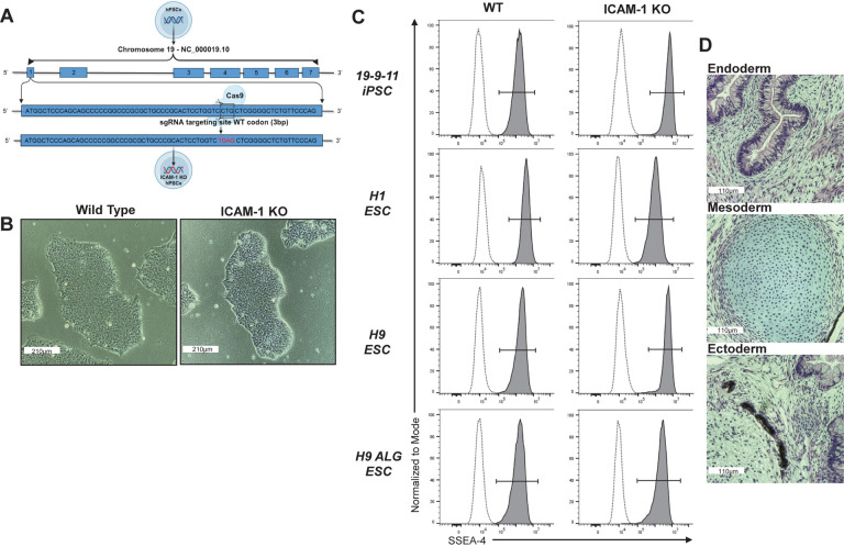
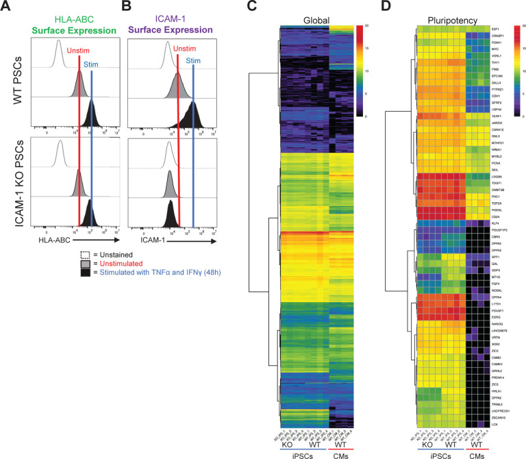
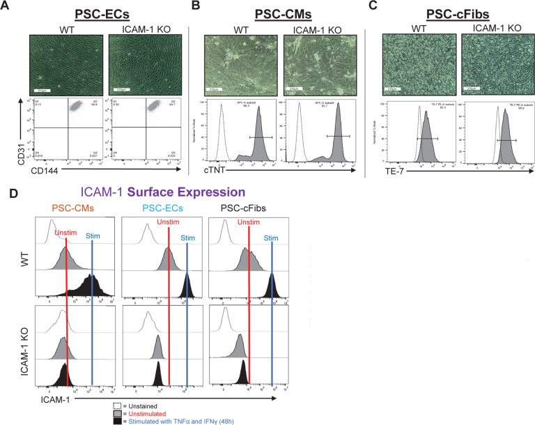
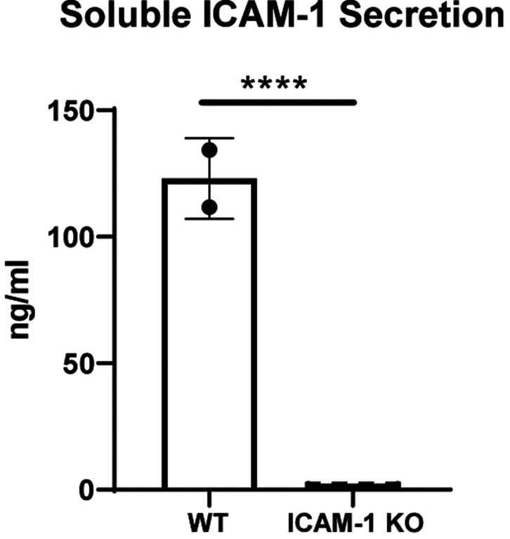
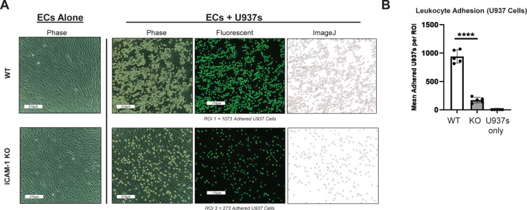
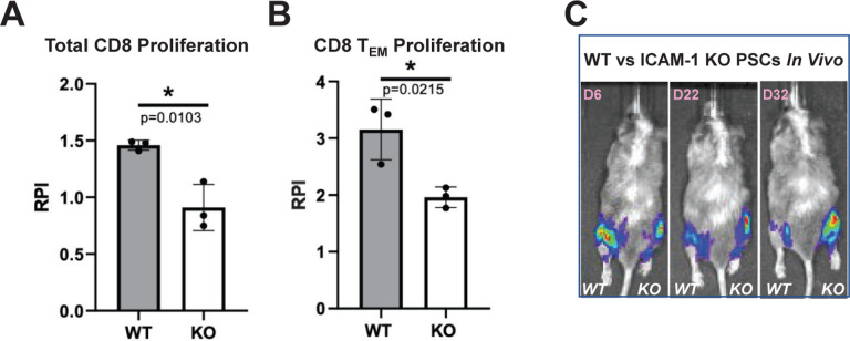
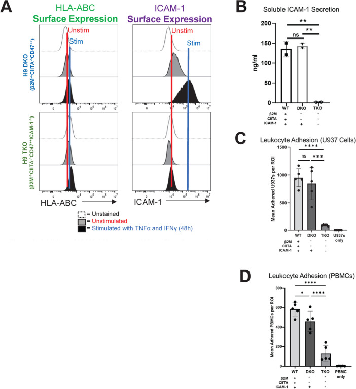
Hypoimmune gene edited human pluripotent stem cells (hPSCs) are a promising platform for developing reparative cellular therapies that evade immune rejection. Existing first-generation hypoimmune strategies have used CRISPR/Cas9 editing to modulate genes associated with adaptive (e.g., T cell) immune responses, but have largely not addressed the innate immune cells (e.g., monocytes, neutrophils) that mediate inflammation and rejection processes occurring early after graft transplantation. We identified the adhesion molecule ICAM-1 as a novel hypoimmune target that plays multiple critical roles in both adaptive and innate immune responses post-transplantation. In a series of studies, we found that ICAM-1 blocking or knock-out (KO) in hPSC-derived cardiovascular therapies imparted significantly diminished binding of multiple immune cell types. ICAM-1 KO resulted in diminished T cell proliferation responses in vitro and in longer in vivo retention/protection of KO grafts following immune cell encounter in NeoThy humanized mice. The ICAM-1 KO edit was also introduced into existing first-generation hypoimmune hPSCs and prevented immune cell binding, thereby enhancing the overall hypoimmune capacity of the cells. This novel hypoimmune editing strategy has the potential to improve the long-term efficacy and safety profiles of regenerative therapies for cardiovascular pathologies and a number of other diseases.


Conflict of interest statement

Conflict of Interest Disclosure M.E.B is a consultant for Taconic Biosciences, and is an inventor on a patent application filed by WARF related to this work. The other authors declare no conflict of interest.

Figures

Figure 1.
Figure 1.. Adhesion Assay with Antibody Blocking of ICAM-1 in Pluripotent Stem Cell (PSC)-Derived Cardiovascular Cells.
(A) The U937 monocytic cell line was stained with LFA-1 (CD11a/CD18) and MAC-1 (CD11b/CD18) alpha chain antibodies. (B) PSCs (H9 embryonic stem cell line) were differentiated into high-purity endothelial cells (ECs) (>85% CD31+CD144+), stimulated with TNFα (10ng/ml) and IFNγ (50ng/ml) for 48 hours, followed by incubation with anti-human ICAM-1 blocking antibody (αICAM-1) (0.5mg/ul) for 1 hour at 37°C, and washed. U937 cells (labeled with fluorescent Calcein AM dye) were then incubated on the ECs for 20 minutes to allow binding, washed to remove any unbound U937 cells, and imaged. Image analysis via ImageJ (version 1.54g) showed binding of U937 cells to target PSC-derived ECs, (C) PSC-derived cardiomyocytes (CMs), and (D) PSC-derived cardiac fibroblasts (cFibs) following antibody blocking compared to unblocked controls. **** = p<0.0001, BR=3. Statistical analysis performed via Prism 10.2.2. ROI = Region of Interest
Figure 2.
Figure 2.. Generation of ICAM-1 Knock-out Pluripotent Stem Cell (PSC) Lines.
ICAM-1 was knocked-out (KO) via CRISPR/Cas9 editing. (A) Schematic demonstrating the CRISPR/Cas9 KO strategy. The wild type (WT) codon (CTG) in the first exon of the ICAM-1 sequence is edited into a stop codon (TGA) via the addition of a nucleotide. (B) Representative PSC colony morphology (10X). (C) Pluripotency marker SSEA-4 staining by flow cytometry. (D) Teratomas were grown in immune-deficient mouse hosts by intramuscular injection of the H9 ICAM-1 KO PSC line with Matrigel. Representative Hematoxylin and Eosin stained images of (top) gut [Endoderm], (middle) cartilage [Mesoderm], and (bottom) retinal pigment epithelium [Ectoderm] representing all three germ layers. Multiple cell lines were made in human embryonic stem cells (hESCs) and in human induced PSCs; data are representative of all lines.
Figure 3.
Figure 3.. Transcriptomic and Protein Expression Analysis of ICAM-1 Knock-out (KO) vs Wild Type (WT) Cells.
ICAM-1 KO and WT isogenic pluripotent stem cells (PSCs) were stimulated with TNFα (10ng/ml) and IFNγ (50 ng/ml) for 48 hours, then assessed for (A) MHC class I expression and (B) surface ICAM-1 expression at baseline and following stimulation. (C) Bulk RNA sequencing was performed on ICAM-1 KO vs. WT induced PSCs (iPSCs); WT iPSC-derived cardiomyocytes (CMs) shown as a differentiated cell type reference control. Log2 RSEM counts for 21,055 transcripts from four cell culture wells (biological replicates, BRs) of PED05 ICAM-1 KO iPSCs, four BRs of PED05 WT iPSCs, and four BRs of PED05 WT iPSC-derived CMs. Clustering was performed and the graphic was made by running pheatmap in R Studio using Euclidean distance and ward.D hierarchical clustering metrics. (D) Log2 RSEM counts for 60 gene reference markers classifying PSC identity.
Figure 4.
Figure 4.. ICAM-1 Knock-out (KO) Pluripotent Stem Cell (PSC) Differentiation Capacity and Surface ICAM-1 Protein Expression.
Morphology of (A) PSC-derived cardiomyocytes (CMs), (B) PSC-derived endothelial cells (ECs), and (C) PSC-derived cardiac fibroblasts (cFibs) (top panels) is shown (10x). The purity of cell-type specific markers was determined by flow cytometry (bottom panels). (D) ICAM-1 surface expression is shown at unstimulated baseline and following 48 hours of stimulation with TNFα (10ng/ml) and IFNγ (50ng/ml). Data are representative of n=3 experiments, with triplicated biological replicates. Analysis was performed via FlowJo 10.10 software. Other abbreviations: WT – wild type.
Figure 5.
Figure 5.. Soluble ICAM-1 Secretion.
Pluripotent stem cells were differentiated into highly pure (>96% CD31+CD144+) endothelial cells and stimulated with 10 ng/ml TNFα and 50 ng/ml IFNγ for 48 hours. Soluble ICAM-1 secreted into the cell culture media was assessed by Luminex assay. Unpaired t test, **** = p<0.0001, n=2 biological replicates for wild type (WT) and n=4 biological replicates, n=3 independent experiments. Statistical analysis was performed via Prism 10.2.2 software. Data are representative of n=3 experiments. Other abbreviations: KO – knock-out.
Figure 6.
Figure 6.. Immune Cell Adhesion Assay with ICAM-1 Knock-Out (KO) Pluripotent Stem Cell (PSC)-Derived Endothelial Cells (ECs).
H9 wild type (WT) and ICAM-1 KO PSCs were differentiated into high-purity CD31+CD144+ ECs and stimulated with TNFα (10ng/ml) and IFNγ (50ng/ml) for 48 hours. As in Figure 1, U937s were co-cultured with both cell types and washed. Images were acquired on ECHO Revolve | R4 microscope. **** p<0.0001, biological replicates=3. Statistical analysis was performed via Prism 10.2.2 software. ROI = Region of Interest
Figure 7.
Figure 7.. In Vitro and In Vivo Immune Responses to ICAM-1 Knock-out (KO) Cells.
To test in vitro immune responses to edited cells, highly pure (>85% cTNT+) PSC-derived CMs were made from wild type (WT) and ICAM-1 KO pluripotent stem cells (PSCs), similar to Figure 1C. Cells were co-cultured for 6 days with HLA-mismatched peripheral blood mononuclear cells (1:6 T:E ratio) labeled with VPD450 proliferation dye in a mixed lymphocyte reaction. Alloreactivity was assessed via proliferation (VPD450 dye dilution) of total viable cells and various T cell subpopulations by flow cytometry. Proliferation of (A) Total CD8+CD3+ T cells and (B) Effector memory T (TEM) cells (CD45RO+ CD45RACD62LCCR7) is shown. Relative proliferation index (RPI) is the ratio of % proliferating cells with cardiomyocyte targets/baseline proliferating cells without targets. N=3 biological replicates, error bars = standard deviation, analysis via 2-tailed unpaired t-test, Prism 9.3.1. N=3 repeat experiments. (C) To test in vivo immune responses to edited cells, NeoThy humanized mice with flow-confirmed hCD45+ and hCD3+ (both >10%) immune cells were engrafted with 1e6 ICAM-1 KO (right leg) vs. WT (left leg) isogeneic PSCs co-injected with Matrigel. Humanized immune systems were HLA-mismatched to the PSC grafts. Bioluminescence imaging (BLI) signal was monitored for 32 days at early (6 days), middle (22 days), and late/terminal (32 days) time points. A representative mouse is shown reflecting loss of WT graft, and retention of KO graft, which was seen in 4 of 5 mice BLI signal quantified by total flux (p/s).
Figure 8.
Figure 8.. Addition of ICAM-1 Knock-out (KO) to First Generation Hypoimmune Pluripotent Stem Cell (PSCs).
(A) “First generation” B2M−/−, CIITA−/−, CD47++ (DKO) PSCs were edited to introduce the ICAM-1-KO, making a triple KO (TKO) cell line. PSCs were differentiated into highly pure (>98% CD31+CD144+) endothelial cells (ECs) and stimulated with 10 ng/ml TNFα and 50 ng/ml IFNγ for 48 hours and assessed for HLA class I (left) and ICAM-1 (right) surface protein expression. (B) Wild type (WT), DKO, and TKO PSCs were differentiated into highly pure (>98% CD31+CD144+) ECs and stimulated as above, then assessed by Luminex for secreted soluble ICAM-1, as in Figure 5. (C) Similar to Figures 1 and 6, adhesion assays were performed using U937s as immune effector cells. U937s were stained with violet VPD450 dye. (D) Adhesion assays were also conducted with peripheral blood mononuclear cells (PBMCs). N=3 biological replicates, error bars = standard deviation, analysis via 2-tailed unpaired t-test. WT = wild-type H9 PSC line with ALG (Akaluc + GFP) reporter construct, DKO = H9 PSC line with KO of β2M and CIITA, with ALG reporter construct, TKO = DKO line with addition of ICAM-1 KO. * p<0.05, ** p<0.01, *** p=0.0001, **** p<0.0001, ns = not significant. Statistical analysis was performed via Prism 10.2.2 software.

Similar articles

References

    1. Yu J. et al. Induced Pluripotent Stem Cell Lines Derived from Human Somatic Cells. Science 318, 1917–1920 (2007). - PubMed
    1. Takahashi K. et al. Induction of pluripotent stem cells from adult human fibroblasts by defined factors. Cell 131, 861–872 (2007). - PubMed
    1. Yan W. et al. Stem cell-based therapy in cardiac repair after myocardial infarction: Promise, challenges, and future directions. J. Mol. Cell. Cardiol. 188, 1–14 (2024). - PubMed
    1. Piao J. et al. Preclinical Efficacy and Safety of a Human Embryonic Stem Cell-Derived Midbrain Dopamine Progenitor Product, MSK-DA01. Cell Stem Cell 28, 217–229.e7 (2021). - PMC - PubMed
    1. Ramzy A. et al. Implanted pluripotent stem-cell-derived pancreatic endoderm cells secrete glucose-responsive C-peptide in patients with type 1 diabetes. Cell Stem Cell 28, 2047–2061.e5 (2021). - PubMed

Publication types